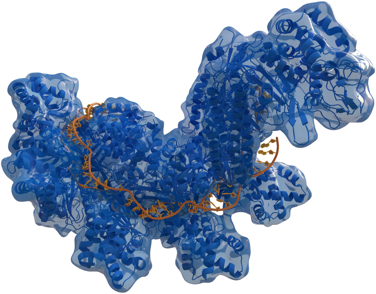
For most of its history, structural biology has worked in still photographs. Researchers determine a protein’s three-dimensional shape and treat that snapshot as the molecule. But proteins are not statues. They flex, breathe and partly unfold, and much of what they do depends on those motions. Capturing them experimentally has been slow and costly, usually managed for only one or a few proteins at a time.
A study published in Nature scales that work up dramatically. Using a new technique, an international team measured the motions of 5,778 protein domains under identical conditions, and found that proteins with nearly the same structure can behave very differently over time.
Hundreds of Proteins in One Experiment
The method combines hydrogen-deuterium exchange with multiplexed mass spectrometry, abbreviated mHDX-MS. Proteins are placed in water containing deuterium, a heavier form of hydrogen. Hydrogen atoms in the protein gradually swap places with deuterium from the solvent. In tightly folded, protected regions the swap happens slowly. In mobile or exposed regions it happens quickly, and those regions gain mass faster. Measuring the rates reveals where a protein flexes and how much energy each local motion requires.
The innovation is doing this for many proteins at once. The domains studied were 28 to 64 amino acids long and drawn from ten protein families. Allan Ferrari, the study’s first author and now a professor of pharmacology at Northwestern Medicine in Chicago, described the approach as mapping the energy landscape of hundreds of molecules in a single experiment, exposing the alternative shapes a protein can adopt beyond its most common one. Ferrari began the project as a postdoctoral fellow at the State University of Campinas in Brazil, supported by the São Paulo Research Foundation, and spent more than five years on it in the laboratory of Gabriel Rocklin, the study’s corresponding author.
Same Shape, Different Behavior
The headline finding is that sequence matters for motion in ways structure alone cannot show. Changing only the amino acid sequence could markedly alter a protein’s dynamics and stability. Proteins within the same structural family sometimes differed from one another more than proteins from different families did. In the most dynamic proteins, whole structural elements, such as helices and sheets, tended to unfold together.
Lysozyme, an enzyme in tears and saliva that breaks down bacterial cell walls, illustrates why this matters for medicine. Certain mutations make it less stable, exposing sticky regions that clump together. The aggregates accumulate in the liver and kidneys and can lead to organ failure. Under conventional structural methods, normal and mutant lysozyme look almost identical. Dynamic mapping shows that the mutation lets part of the structure open more easily.
Techniques such as crystallography, NMR and cryo-electron microscopy often miss brief changes of shape like these, Ferrari noted, and they require each variant to be studied separately.
A Movie for AlphaFold
The work arrives at a moment when artificial intelligence has transformed structure prediction. AlphaFold, trained on more than 50 years of structures in the Protein Data Bank, can predict a protein’s shape from its sequence, and its developers shared the 2024 Nobel Prize in Chemistry. But AlphaFold predicts the snapshot, not the movie. Until now, no large dataset had systematically described protein dynamics to train such models.
The team has released its measurements as the first publicly available experimental database of large-scale protein dynamics. “There’s an unlimited number of possible combinations of amino acids,” Rocklin said. Designing drugs or biosensors, he explained, means knowing which sequence performs best and how changes alter its behavior, and the new method makes it possible to examine those fluctuations across thousands of sequences.
As a test of the data’s usefulness, the researchers chose two mutations predicted to stabilize a naturally flexible region of one protein. Follow-up experiments confirmed the effect.
Linking sequence to transitions between protein states remains an open problem, and the domains studied here are small compared with many proteins in the body. Ferrari plans to generate more data for specific protein families and to study how changes in dynamics contribute to human disease, including how a single mutation can destabilize a protein enough to cause harm without unfolding it entirely.
Endnotes
- Ferrari, A., et al. (May 13, 2026). “Large-scale discovery, analysis and design of protein energy landscapes.” Nature. https://doi.org/10.1038/s41586-026-10465-z
- São Paulo Research Foundation (FAPESP) (August 10, 2026). “New technique reveals on a large scale how thousands of proteins change shape and move.” EurekAlert! https://www.eurekalert.org/news-releases/1139638
Featured image: Structure of the Cascade protein complex, shown as an example of a protein’s three-dimensional form. Credit: Thomas Splettstoesser.


Leave a Reply