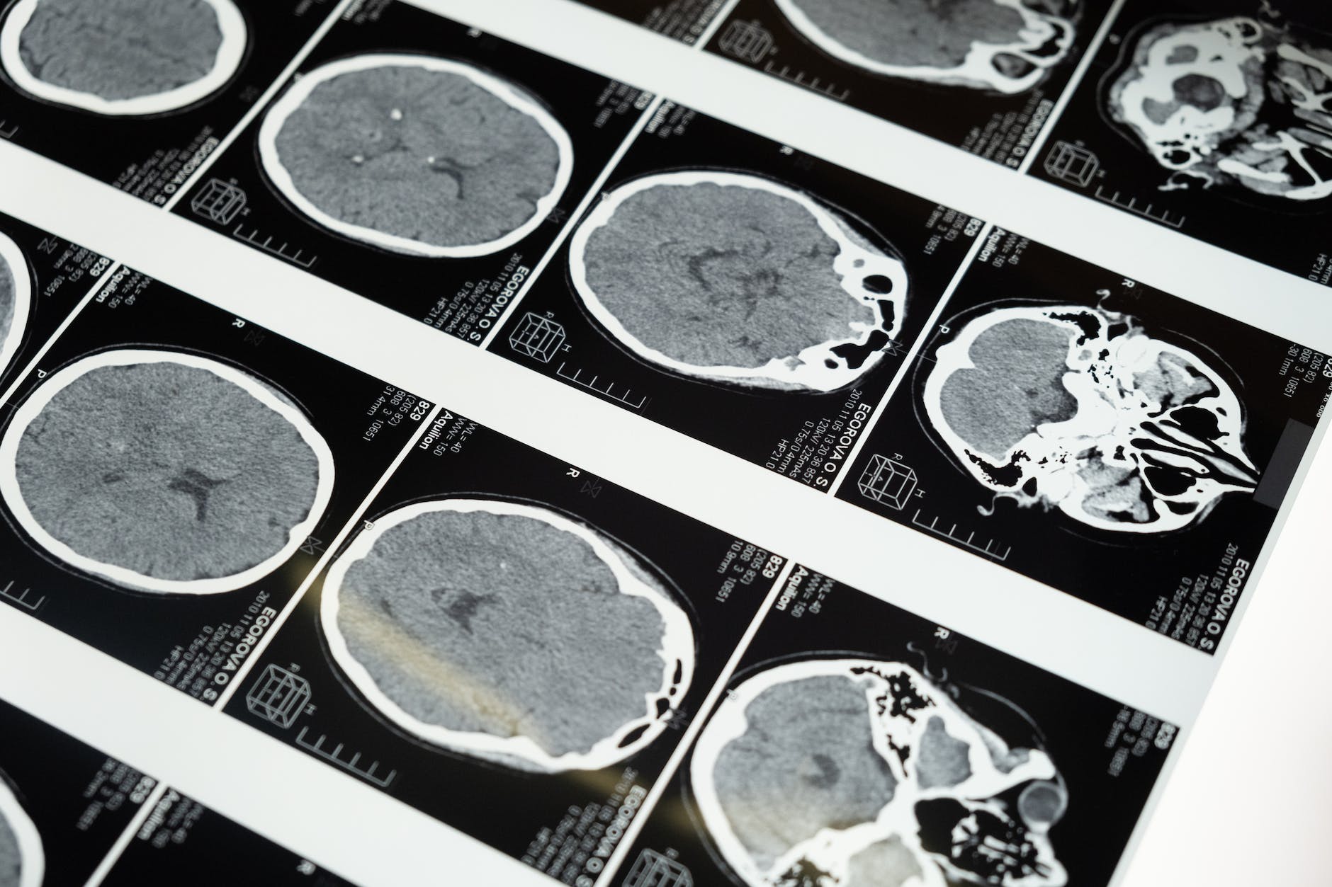
An MRI study of 61 women with breast cancer and 41 healthy women found that fatigue more than doubled after two chemotherapy cycles, tracking changes in a region called the left isthmus cingulate cortex.
Among the side effects of cancer treatment, fatigue is one of the most common and one of the hardest to explain. It is not the tiredness of a long day. Patients describe a deep exhaustion that rest does not relieve, and it can persist for months or even years after treatment ends.
A new study suggests that the brain may begin to change in ways linked to this fatigue very early in treatment. Writing in the journal Cancer, a team led by Professor Li Hai of the Hefei Institutes of Physical Science, part of the Chinese Academy of Sciences, reports brain changes associated with cancer-related fatigue in women with breast cancer after just two cycles of chemotherapy.
A common and stubborn problem
Cancer-related fatigue is thought to affect a majority of people undergoing chemotherapy at some point. It can make it hard to work, care for family or keep up with daily activities, and it is a frequent reason for reduced quality of life during and after treatment.
Its causes are not well understood. Researchers have pointed to inflammation, anemia, disrupted sleep, emotional distress and the direct effects of cancer and its treatments on the body and brain. Many patients also report cognitive problems during chemotherapy, sometimes called “chemo brain,” suggesting that the brain itself can be affected.
Breast cancer is the most commonly diagnosed cancer in women worldwide, and many patients receive chemotherapy before or after surgery. Because survival rates have improved substantially, a growing number of people are living for years with the after-effects of treatment, making problems such as fatigue an increasingly important focus of research.
Scanning before and during treatment
To look for early brain changes, the researchers followed 61 women with breast cancer and 41 healthy women. All of them underwent brain MRI on a 3-tesla scanner, along with clinical assessments, before chemotherapy began and again after two treatment cycles. The healthy women were scanned at matching times.
Over those two cycles, the proportion of patients with clinically significant fatigue rose from 29.5 percent to 70.5 percent.
The team measured two aspects of the cerebral cortex, the brain’s outer layer. One was cortical thickness, a standard measure of brain structure. The other was cortical mean diffusivity, which tracks how freely water molecules move within tissue and can reveal subtle changes in the microscopic structure of the cortex before they show up as changes in thickness.
Several cortical regions showed changes in both measures.
Diffusion measures are sensitive to the microscopic environment of tissue, such as the density of cells and the space between them. A drop in mean diffusivity means water is moving less freely, which can reflect changes such as swelling of cells or shifts in tissue structure, although the measure alone cannot say which process is responsible.
One region stands out
The most notable change was in the left isthmus cingulate cortex, part of a band of tissue that curves around the middle of the brain. There, mean diffusivity fell more sharply in patients than in healthy women over the study period.
The change was linked to symptoms. Patients whose mean diffusivity fell more also showed greater worsening of fatigue and of emotional well-being.
The isthmus cingulate cortex lies in the posterior cingulate region, which is part of the brain’s default mode network, a set of regions most active when people are at rest or turning attention inward. The region has been linked to self-referential thought, memory and emotional processing.
The researchers’ analysis suggested that changes in emotional well-being may partly explain the connection between the brain changes and fatigue. In other words, the brain change may affect mood, which in turn contributes to exhaustion, although the study cannot prove that sequence.
What the findings mean
The authors conclude that brain changes related to fatigue can appear early during chemotherapy, and that the left isthmus cingulate cortex may play a role in both fatigue and emotional well-being.
If confirmed, such early markers could help identify patients at risk of severe fatigue and point toward ways to intervene before symptoms become entrenched.
The study has limitations. It was relatively small, followed patients only through the first two cycles of treatment and involved only women with breast cancer, so the findings may not apply to other cancers or treatments. The release does not describe the chemotherapy regimens used, and changes in diffusion measures can reflect many different processes in tissue, so what is happening at the cellular level remains unclear. The release also does not list the study’s full authorship or funders.
Notes
- “Early neural signatures of cancer-related fatigue: A longitudinal magnetic resonance imaging study of cortical micro- and macrostructural changes during breast cancer chemotherapy.” Cancer, 28 July 2026. DOI: 10.1002/cncr.70558.
- Hefei Institutes of Physical Science, Chinese Academy of Sciences. “Brain changes linked to fatigue emerge early during chemotherapy.” EurekAlert!, 1 August 2026. https://www.eurekalert.org/news-releases/1141892.
Photo by cottonbro studio on Pexels.com

Leave a Reply