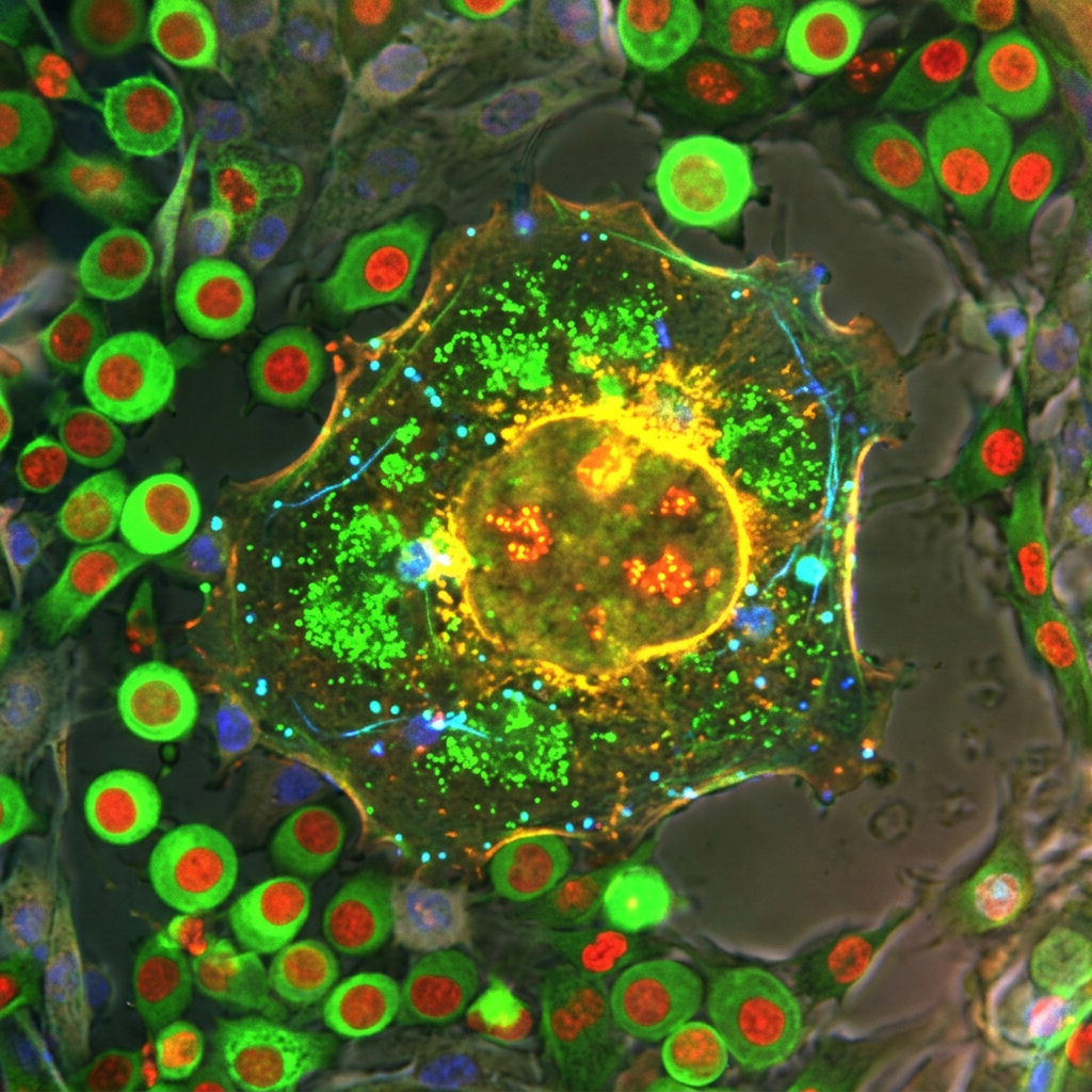
Eat more protein. Cut back on sugar. Get more fibre. Nutrition advice is everywhere, and nearly all of it rests on a surprisingly thin foundation. Scientists have only a limited view of what any particular nutrient does once it reaches an individual cell.
Tara TeSlaa, an assistant professor of molecular and medical pharmacology at the University of California, Los Angeles, and a member of the UCLA Broad Stem Cell Research Center, has been awarded an NIH Director’s New Innovator Award to change that. The grant, which supports early-career scientists pursuing unusually innovative work, provides up to $2.375 million in direct research costs over five years. TeSlaa plans to use it to map how different types of cells use nutrients inside the body.
The gap she is addressing helps explain why nutrition studies so often seem to contradict one another. Research frequently links a nutrient to a health outcome without identifying the cellular mechanism behind it. Take the conflicting literature on dietary fat. “What’s good for one cell type may not necessarily be good for another,” TeSlaa said.
A Measurement Problem
Cell metabolism is a moving target. Cells change which nutrients they draw on as supply shifts with diet, disease, exercise and other changes in the body, and those shifts can alter how the cells behave. The molecules that record this activity, called metabolites, are the small compounds a cell uses or produces as it processes nutrients. Many of them last only seconds or minutes.
That is far less time than it takes to isolate a specific cell type. Doing so means breaking tissue apart, a slow process that also pulls cells out of their natural surroundings and may change the very metabolism a researcher is trying to measure.
Growing cells in a dish avoids some of those problems and introduces another. Cultured cells can use nutrients differently from the way they do in the body. Cancer cells grown in the laboratory, for example, consume more glutamine and less glucose than the same cells inside a tumor. That discrepancy may be part of the reason some cancer drugs aimed at metabolism look promising in the lab and then fail in animal models. And because cell-specific measurements cannot routinely be made in living people, researchers depend on models that may not fully reflect what happens in a body.
Writing Metabolism Into Longer-Lasting Molecules
TeSlaa’s answer is to make cells keep their own records. Her lab calls the method STOMP, for Stable isotope Tracing of Orthogonal Metabolites into bioPolymers. Researchers supply nutrients tagged with stable, nonradioactive isotopes. As cells process those nutrients, the tags pass into metabolites and from there into longer-lived molecules such as proteins and DNA.
Because proteins and DNA are assembled over time and persist, they preserve a durable account of what the cell consumed while it was still in place. The cells can then be isolated at leisure and the record read with mass spectrometry, which detects the isotope labels. The team has already shown the approach can work. The award is meant to let them scale it up.
The first targets are the liver and the pancreas, both central to metabolic conditions such as fatty liver disease and diabetes. The liver regulates metabolism across the whole body. The pancreas contains the beta cells that sense blood glucose and release insulin, but those cells make up a small fraction of the organ and are slow to isolate, which has made their metabolism in the body hard to study.
Rare Cells and Imperfect Models
A map drawn at the level of cell types could bring rare but important cells into view. In the liver, hepatocytes handle most of the major metabolic work, and measurements of whole tissue mostly reflect them. Less abundant cells, including immune cells and hepatic stellate cells, can drive inflammation and scarring after the liver is damaged. TeSlaa wants to learn how those cells respond to changes in diet or disease. Eventually she plans to extend the method to the brain, heart, skeletal muscle, spleen, kidney, intestine, skin and fat.
The project should also show how faithfully laboratory stand-ins reproduce metabolism in the body. Researchers often rely on tissue slices and on organoids grown from stem cells. TeSlaa plans to compare those systems with measurements from mouse models to see which best preserve natural metabolic behavior.
That question is sharpest for tissue stem cells, which are scarce and can change their behavior as soon as they are removed. Her lab is particularly interested in muscle stem cells, which lie mostly dormant until a muscle needs repair. Recording their metabolism before removal could reveal how nutrient use shifts as the cells wake up and rebuild tissue, and how aging or exercise change the pattern.
This is an award announcement, not a report of results, and the map does not yet exist. TeSlaa describes it as a starting point for understanding metabolism one cell type at a time.
Endnotes
- University of California, Los Angeles Health Sciences (October 6, 2026). “What do your cells actually eat?” EurekAlert! https://www.eurekalert.org/news-releases/1146740

Leave a Reply