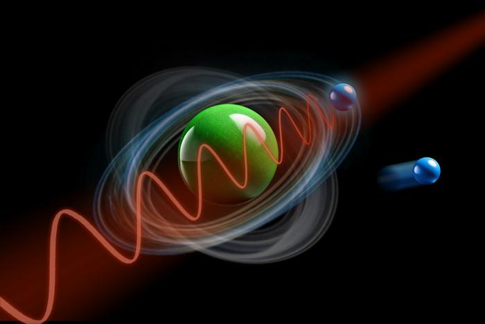
Göttingen physicists imaged a molecular orbital’s three-dimensional shape with a laboratory soft-X-ray laser, a measurement that until now needed a synchrotron.
The wavefunction is the central object of quantum mechanics and the one you cannot point a camera at. It is not a thing in space but a complex-valued function whose squared magnitude gives the probability of finding an electron somewhere, and the measurement problem is that any measurement collapses it.
“The wavefunction is a fundamental quantity in quantum mechanics, yet it cannot be directly observed or measured,” said Professor Stefan Mathias of the Faculty of Physics at the University of Göttingen, who leads the Ultrafast Dynamics in Quantum Materials research group.
What can be done is reconstruction, and a paper in Nature Communications reports doing it in three dimensions for a nanometre-sized organic molecule, using a light source that fits on a laboratory table.
How you reconstruct something you cannot see
The technique is photoemission orbital tomography, and the physics behind it is a useful piece of luck. Shine high-energy light on a molecule and an electron is ejected; the angular distribution of those photoelectrons is related to the spatial distribution of the orbital the electron came from, approximately by a Fourier transform.
Measure the photoelectron intensity across enough angles and energies and the orbital’s shape can be computed back out. Doing it in two dimensions has been established for over a decade. Doing it in three requires sampling the photoelectron momentum distribution over a volume rather than a plane, which means many more measurements.
That is why the three-dimensional version has previously been the province of synchrotrons — large shared facilities where the required photon energies are available and where beam time is scarce and the measurements are slow.
Two changes
The Göttingen work removes the facility requirement by attacking the problem from both ends: fewer measurements needed, and a light source that can sit in a university laboratory.
“We introduce two powerful new concepts. First, by redesigning the computer algorithm from the ground up, reliable 3D images can now be obtained,” said Dr Matthijs Jansen, who co-led the work.
The second is the source: a femtosecond extreme-ultraviolet light source producing ultrashort pulses, rather than a synchrotron beamline. The reconstruction resolved features smaller than the spacing between the molecule’s own carbon atoms, which is the relevant benchmark — a reconstruction coarser than the bond lengths would not tell you anything about orbital structure.
The algorithmic half is doing more work here than it might appear. Reducing the data a reconstruction needs is what converts a measurement from a facility campaign into a laboratory experiment, and it is a change in what questions can be asked rather than only in where they are asked.
There is an applied reason to want that. Organic semiconductors — the materials in OLED displays and in printable solar cells — work by moving charge between molecular orbitals, and how well they work depends on the shapes of those orbitals and how they overlap between neighbouring molecules. Those shapes are currently predicted computationally and checked indirectly, and a measurement that can be run in a materials laboratory rather than booked at a national facility is the difference between validating a calculation occasionally and doing it routinely.
Towards watching an orbital change
The reason to care about ultrashort pulses is that they open the time domain. A femtosecond is a quadrillionth of a second, which is the timescale on which electrons rearrange during chemical reactions, and a source that delivers pulses that short can in principle take snapshots at intervals that short.
“This technique might mean that stroboscopic videography becomes a reality,” said Wiebke Bennecke, the paper’s first author — allowing the shape of a wavefunction to be followed as it changes rather than recorded once.
That would be a genuinely different kind of observation. Most of what is known about electronic structure during a reaction is inferred from spectroscopic signatures and compared against calculation; a sequence of reconstructed orbitals through a bond breaking would be a direct measurement of the thing the calculations are modelling.
It is also an aspiration rather than a result. The paper demonstrates a static three-dimensional reconstruction with a source capable of femtosecond pulses; the time-resolved version has not been done, and the data requirements for reconstructing an orbital at each of many time delays are the obvious obstacle.
What “photographed” is doing in the headline
The release describes the orbitals as photographed in 3D, and the gap between that word and the measurement is worth being clear about.
Nothing was imaged in the sense of light reflecting off an object onto a sensor. Photoelectrons from many identical molecules, over many pulses, were counted as a function of angle and energy, and an algorithm inverted that distribution into a spatial structure. The result is a reconstruction whose fidelity depends on the inversion, on the approximations relating photoelectron angle to orbital shape, and on the molecules being ordered enough that their signals add coherently.
Nor is the full wavefunction recovered. A wavefunction has amplitude and phase, and what these techniques principally determine is the spatial distribution; the phase information is partly reconstructed and partly supplied by the assumptions in the algorithm. “Electron orbital” is the honest description of what comes out, which is the term the paper itself uses.
The release also gives no numbers of any kind beyond the molecule being nanometre-sized and the pulses femtosecond-long. It does not quantify how much the data requirement fell, which is the paper’s headline claim, nor state what molecule was measured, nor report the achieved spatial resolution as a figure. For a methods advance, those are the numbers a reader would want.
The paper was published on 19 June 2026. The release does not state the study’s funding sources, the full author list, or any conflicts of interest.
Notes
- Bennecke, W., Jansen, M., Mathias, S., et al. “Table-top three-dimensional photoemission orbital tomography with a femtosecond extreme ultraviolet light source.” Nature Communications, 2026. DOI: 10.1038/s41467-026-74308-1.
- University of Göttingen. “Zooming in: Electron orbitals photographed in 3D.” EurekAlert!, 4 August 2026. https://www.eurekalert.org/news-releases/1138815.
TU Wien


Leave a Reply