When tissues are held under tension for hours, their keratin networks slowly reorganize into star-shaped bundles that span many cells. In the process, the nucleus is pushed out of its protective cage.
Most of what we know about how living tissue handles force comes from what happens in the first seconds and minutes: a cell is poked, pulled, or squeezed, and it responds. But many tissues in the body don’t experience force as a sudden event. They live under it. The skin over a growing belly, the wall of a filling bladder, the expanding tissue of a mammary gland — these are stretched and held, sometimes for hours, sometimes for days.
How cells cope with that kind of sustained load has been surprisingly murky. A new study published in Nature Physics offers an answer that is both elegant and a little strange. Led by researchers at the Institute for Bioengineering of Catalonia (IBEC), the work shows that epithelial cells under prolonged stretch gradually remodel their internal keratin scaffolding into thick bundles that link neighboring cells into a single, tissue-wide network. As that happens, the nucleus — normally nestled inside a mesh of keratin filaments — is slowly squeezed out of its cage.
A slow-motion response
Keratin is best known as the stuff of hair and nails, but inside cells it forms part of the cytoskeleton, the web of protein filaments that gives cells their shape and lets them resist deformation. Biologists have long understood that keratin helps tissues survive large strains. What they hadn’t pinned down was how the network itself behaves when the strain doesn’t let up.
To find out, the team built engineered sheets of epithelial cells and placed them in a custom microfluidic device capable of stretching the tissue in a controlled way. They tracked the cells with live microscopy and paired the experiments with detailed computational models that simulated the physics inside each cell.
The first surprise was patience. Keratin didn’t respond right away. Only over several hours did individual filaments begin to reorganize, collecting into thick, star-shaped bundles. Those bundles didn’t stay confined to single cells; they reached across cell boundaries, forming what the researchers describe as supracellular networks running through the tissue.
The remodeling starts in a specific place: the points where three cells meet. Keratin filaments gradually vanish from these tricellular junctions and accumulate instead in the growing bundles. And the process spreads. It begins in a handful of isolated cells, then propagates to their neighbors, producing expanding clusters of bundled cells stitched together by a shared keratin framework.
The model that predicted an escape
The most striking result emerged first in the simulations. Marco Pensalfini, an assistant professor at Queen Mary University of London, developed the computational model with Marino Arroyo of the Universitat Politècnica de Catalunya and the International Centre for Numerical Methods in Engineering (CIMNE).
“Simulating the intracellular dynamics revealed that the same process that forms these striking bundles also generates forces that ultimately push the nucleus out of its cage,” Pensalfini said. “Computational modelling allowed us connect what we observed in experiments with the underlying physical mechanisms.”
The team then went looking for that predicted behavior in living tissue — and found it. Live imaging showed that as the keratin bundles thickened, the nucleus progressively detached from the surrounding filament mesh until it separated almost completely, tethered by only a residual thread of keratin. The researchers call this “nuclear uncaging,” and they found it to be tightly linked to bundle formation itself rather than a separate event.
Protection or exposure?
What uncaging means for the cell is an open question, and the researchers are candid that it could cut either way.
“One of the most intriguing questions raised by our study is whether this uncaging process ultimately protects the nucleus or makes it more vulnerable,” said Xavier Trepat, an ICREA research professor at IBEC and co-lead author of the study. “On the one hand, removing the keratin cage could expose the nucleus more directly to mechanical forces. On the other, and perhaps counter-intuitively, disconnecting the nucleus from a highly stressed cytoskeletal network may actually protect it by preventing force transmission.”
That tension matters. The nucleus houses the genome, and mechanical forces transmitted to it are known to influence gene expression and, in extreme cases, damage DNA. A cell that cuts the line between a strained cytoskeleton and its nucleus might be insulating its most precious cargo. Determining which effect wins out, Trepat said, will require future studies of how nuclei fare under long-term mechanical stress.
Actin’s restraining hand
The study also found that keratin doesn’t act alone. Actin, another major cytoskeletal filament, turned out to be a key regulator of the process. When the researchers weakened the molecular connections between actin and keratin, bundling sped up nearly threefold. The interplay between the two filament systems, in other words, sets the pace at which tissues adapt to being stretched.
From bladders to disease
Tom Golde, a postdoctoral researcher in Trepat’s group and the study’s first author, sees relevance well beyond the lab dish. “These findings may help us better understand how tissues adapt to prolonged stretching in a wide range of biological contexts,” he said. “Similar mechanical conditions occur during embryonic development and in organs that repeatedly expand and contract, such as the bladder or the mammary gland. We also think that the mechanisms we uncovered could be relevant to diseases in which the keratin network is altered.”
That last point is significant. Mutations in keratin genes underlie a range of disorders, including skin-blistering conditions in which tissues tear under ordinary mechanical stress. Understanding how healthy keratin reorganizes under load could clarify what goes wrong when it can’t.
The researchers draw an engineering analogy: just as bridges and buildings are reinforced to withstand repeated loading, cells appear to continually rebuild their internal scaffolding to stay resilient. The difference is that a cell does it on its own, slowly, from the inside — and in doing so, it may be making a quiet decision about how much of the world’s force its nucleus should feel.
The work involved collaborators at IBEC, Queen Mary University of London, the Universitat Politècnica de Catalunya, CIMNE, Columbia University, the University of Barcelona, the University of Vienna, and University College London.
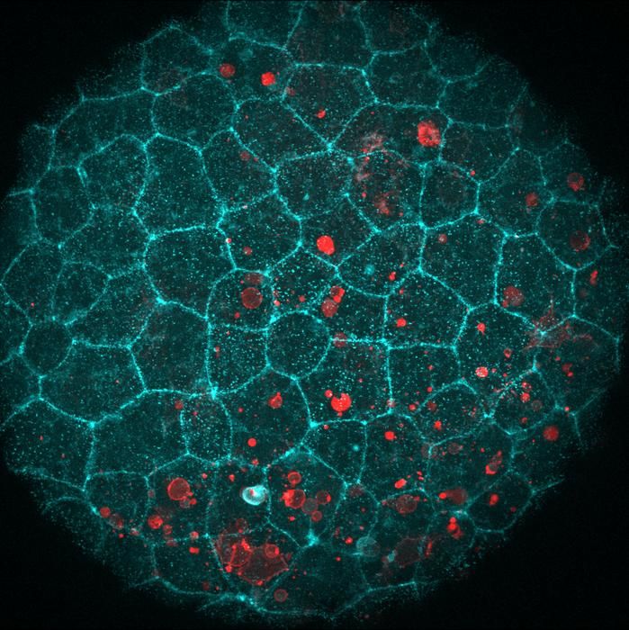
Featured image: A living zebrafish embryo with actin shown in cyan. Credit: Laura F. Bianchi/Centro de Regulación Genómica
Notes
- Golde, T., et al. Study on keratin reorganization and nuclear uncaging under sustained stretch. Nature Physics, July 27, 2026. DOI: 10.1038/s41567-026-03371-8.
- Queen Mary University of London. “Under pressure: When tension builds up, the nucleus escapes.” News release via EurekAlert!, July 27, 2026. https://www.eurekalert.org/news-releases/1137663


Leave a Reply