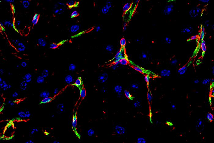
Live imaging in zebrafish reveals how cells link up and crawl forward to join separate vessel segments into one continuous tube, and why a single molecule does double duty in the process.
The human body contains a vascular network so extensive that, laid end to end, it would wrap around the planet more than once. Every one of those vessels had to be built, cell by cell, into a hollow tube with an uninterrupted channel through which blood could flow. Get it wrong, and tissue starves. Get it right, and every organ receives the oxygen and nutrients it needs.
How cells pull off that feat has been only partly understood. Now researchers at the University of Basel’s Biozentrum have watched it happen in remarkable detail. Using high-resolution live imaging in zebrafish, a team led by Professor Markus Affolter and Dr. Heinz-Georg Belting has documented, for the first time, the complete sequence by which separate vessel channels connect and merge into one continuous blood vessel. The study appears in the journal eLife.
The answer turns out to be mechanical. Cells push, anchor themselves to their neighbors, and pull, repeating the cycle again and again like an inchworm working its way along a branch.
The problem of building a tube
Blood vessels are lined by endothelial cells, which form the inner wall of every artery, vein and capillary. As vessels develop, these cells must reshape themselves and rearrange their positions to create hollow tubes, each with a continuous interior channel, called a lumen.
The challenge is that the cells must do this while staying connected to one another. They are joined by cell-cell junctions, structures that hold neighboring cells together. Those junctions face competing demands. They need to be stable enough to keep the vessel wall intact and leak-free, yet flexible enough to let cells slide past and around each other as the vessel takes shape.
The Basel team had previously shown that specialized membrane protrusions called junction-based lamellipodia, or JBL, form the leading edge of endothelial cells and drive their rearrangement. What remained unclear was which physical forces allowed the cells to stretch out and connect.
Watching cells crawl
Zebrafish are a favorite model for this kind of work. Their embryos are transparent and develop outside the mother’s body, so researchers can watch blood vessels form in a living animal under a microscope. By imaging those vessels at high resolution, the Basel team could follow individual cells as they moved.
What they saw was a repeating three-step cycle. First, a JBL protrusion forms at the tip of an endothelial cell and pushes outward, extending the cell forward. At its very tip, the protrusion acts as a molecular anchor, latching onto the neighboring cell. That anchor links the two cells together and stabilizes the developing vessel.
Then pulling forces take over, drawing the rear of the cell forward and gradually stretching it out. With each cycle, the cell advances and the lumen extends a little further.
“Through this repeated cycle of pushing and pulling, the cells move forward much like an inchworm, progressively extending the lumen,” explained Dr. Ludovico Maggi, the study’s first author. Through this process, segments that started out separate eventually merge into a single vessel with an unbroken channel.
One molecule, two jobs
At the heart of the process is a protein called VE-cadherin, which is found at the junctions between endothelial cells. It has long been known as the adhesive that holds those cells together. The new work shows it does considerably more.
“We found that the molecule VE-cadherin performs two distinct functions,” said Belting, the study’s last author. “It not only acts as the ‘glue’ that holds endothelial cells together and maintains the stability of the vessel wall but also plays an active role in cell movement.”
That dual role helps resolve the apparent paradox of junctions that must be both stable and dynamic. The same molecule that keeps the vessel wall sealed is also part of the machinery that lets cells migrate and rearrange.
The behavior of the cells themselves was also unexpected. “We were also surprised by how remarkably plastic these cells are,” Belting said. “Both, stable and highly flexible at the same time.”
More than plumbing
The study provides the first detailed account of how endothelial cells coordinate their junctions during vessel formation and demonstrates that both pushing and pulling forces are essential to producing continuous vessels. Its significance extends beyond the vascular system itself.
Nearly every organ in the body develops in close partnership with its blood supply. Vessels deliver oxygen and nutrients to growing tissues, but they also send signals that help shape how organs form. Understanding how vessels are built therefore illuminates organ development more broadly.
“Blood vessels are far more than simple supply lines,” Belting said. “They accompany the development of almost every organ. Understanding how blood vessels form means understanding a fundamental aspect of organ formation.”
Toward vascularized organoids
The findings may eventually prove useful in the lab. Researchers increasingly grow organoids, small three-dimensional tissue structures that mimic aspects of real organs, to study disease, test drugs and explore regenerative therapies. One persistent limitation is that most organoids lack functioning blood vessels. Without a vascular supply, they cannot grow beyond a modest size, because cells at their core are starved of oxygen and nutrients, and they fail to replicate the way real organs develop alongside their vessels.
A detailed understanding of the forces and molecules that drive vessel formation could help scientists engineer organoids and other lab-grown tissues with working vasculature. That, in turn, could make them far more faithful models of human organs and bring regenerative medicine a step closer to building functional replacement tissue.
The vision of fully vascularized lab-grown organs remains a long-term goal. But it rests on exactly the kind of fundamental knowledge the Basel study provides: a frame-by-frame account of how cells, through a patient rhythm of pushing and pulling, build the channels that keep the body alive.
Featured image: Sarah Lutz
Endnotes
- Maggi, L., et al. “Junctional and Actomyosin Dynamics Drive Endothelial Cell Rearrangements during Vascular Tube Formation.” eLife (2026). DOI: 10.7554/eLife.109264.2.
- University of Basel. “Blood must flow: How pushing and pulling forces shape our blood vessels.” News release via EurekAlert!, July 28, 2026. https://www.eurekalert.org/news-releases/1137714


Leave a Reply