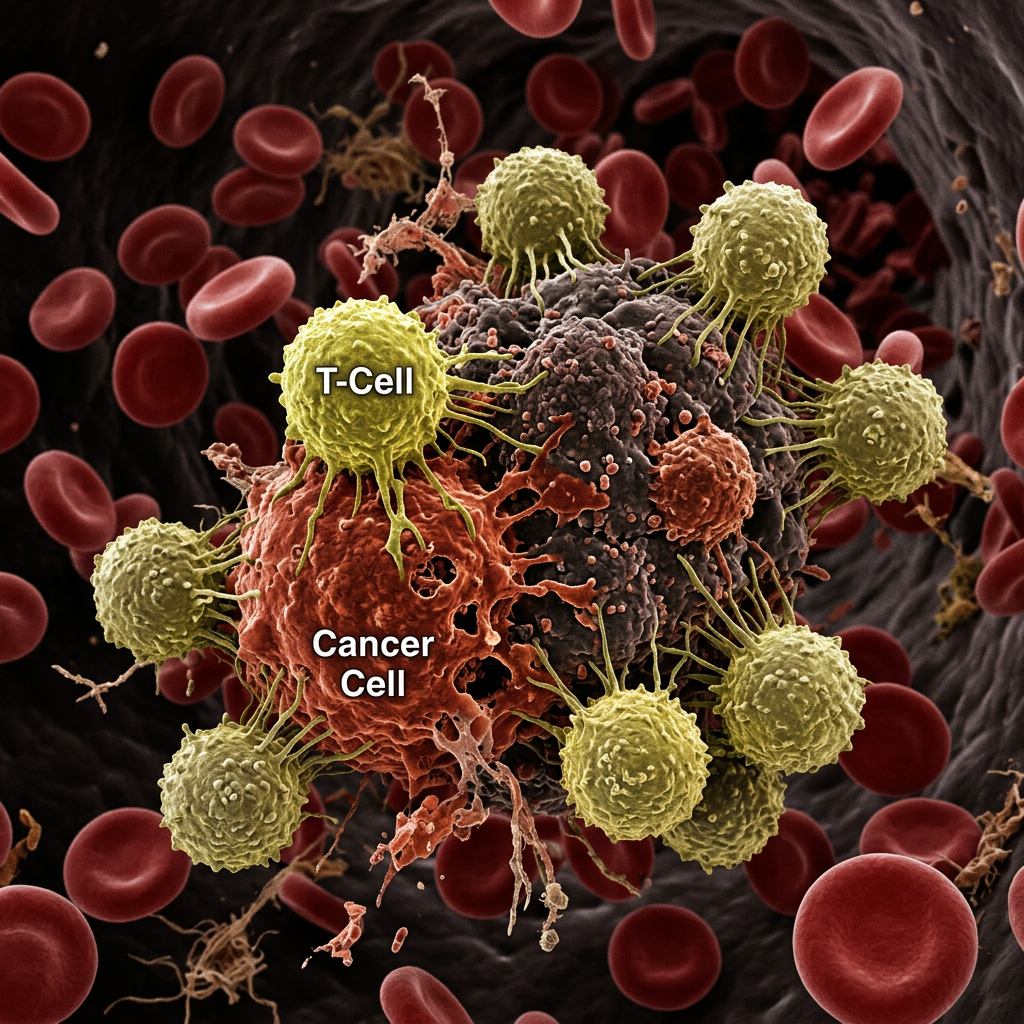
Tohoku University researchers say they have solved nanoparticle clearance and deep-tissue laser delivery at the same time. In mice with colon cancer, a single treatment made tumors vanish within two weeks.
Photothermal therapy has always had an appealing premise. Instead of cutting out a tumor or flooding the body with chemotherapy, doctors deliver light-absorbing particles to cancer cells and then shine near-infrared laser light on them. The particles heat up and the cancer cells cook, while healthy tissue is largely spared. It is far less invasive than surgery or radiation, and it avoids many of the punishing side effects of chemotherapy.
Yet the approach has remained on the margins of cancer care, held back by two stubborn obstacles. The immune system tends to recognize and clear away the nanoparticles before they reach the tumor. And laser light shone from outside the body loses energy as it passes through skin and tissue, making deep tumors hard to heat. A team led by Eijiro Miyako at Tohoku University in Japan now reports that it has tackled both at once.1
“Nanoparticle clearance and laser delivery have long stood in the way of photothermal cancer treatment but we have now overcome both challenges simultaneously,” said Miyako, a professor at Tohoku’s Institute of Multidisciplinary Research for Advanced Materials, who led the work with collaborators at Air Water Inc., Teikyo University, and the Japan Advanced Institute of Science and Technology.2
A protein redesigned by AI
The first problem is a matter of disguise. Nanoparticles injected into the bloodstream are, from the immune system’s point of view, foreign debris to be removed. The standard workaround is to coat them in polyethylene glycol, or PEG, a polymer that helps them slip past immune surveillance for a while. But PEG-coated particles still get cleared relatively quickly.
Miyako’s team turned to AlphaFold, the AI tool that predicts protein structures, to redesign human serum albumin, a protein that circulates naturally in the blood and can help materials evade detection. The result was a new biodegradable protein they call IDP1, which is flexible and water-attracting. In mice, nanoparticles wrapped in IDP1 stayed in circulation for 150.5 minutes, compared with 17.8 minutes for PEG-coated particles — more than four times longer.2
The researchers loaded these IDP1-coated nanoparticles with carbon nanohorns, tiny cone-shaped carbon structures, and indocyanine green, a light-activated dye already used in medical imaging. The resulting particles accumulated efficiently in tumors while showing minimal spread to healthy organs.2
Bringing the light inside
The second problem is one of physics. Near-infrared light penetrates tissue better than visible light, but not infinitely well. Shine a laser on the skin above a deep tumor and much of its energy is absorbed or scattered before it arrives.
The team’s answer was to stop shining the light from outside. Working with graded-index plastic optical fiber lens technology developed by Air Water Inc., they built an ultra-thin rigid endoscope small enough to fit inside a standard 16-gauge needle. The fiber lens was not an adaptation of an existing system, the researchers note, but was designed specifically to match clinical needle sizes.2
Inserted directly into a tumor, the endoscope delivers laser light from within, sidestepping the losses that come from passing through healthy tissue. The system heated tumors as effectively as conventional external treatment while using 28 percent less laser power — 500 milliwatts instead of 700 — which also meant less collateral damage to surrounding tissue. The device includes a real-time fluorescence imaging system, so clinicians can see the tumor and position the endoscope accurately before switching on the laser.2
Tumors gone in two weeks
The combined approach was tested in mice with colon cancer. A single dose of the IDP1-coated nanoparticles followed by one laser treatment caused tumors to disappear completely within 14 days. The tumors did not return during the 40-day study period, and the researchers found no signs of harmful side effects.2
Mouse results are a long way from a clinical therapy, and the team is clear that safety studies come next. But the two components of the work have implications beyond this particular treatment. The AI-guided strategy used to design IDP1 could, in principle, be applied to extend the circulation time of many other drugs that the body clears too quickly. And the needle-sized endoscope could be used to treat other tumors that can be reached with a needle.2
Next targets
Looking ahead, the researchers plan to test the IDP1 platform against cancers that are accessible by endoscope, including head and neck, esophageal, and pancreatic cancers.2 Pancreatic cancer in particular is notoriously difficult to treat and is often diagnosed late, making any minimally invasive option worth watching.
The study was published as an invited contribution to the journal Small Science, where Miyako also serves on the editorial advisory board.1 If the approach survives the move from mice to people, photothermal therapy may finally get the chance its early promise suggested — not by finding a better laser or a better particle alone, but by fixing both problems together.
Notes
- Intrinsically Disordered Polypeptides-Based Stealth Materials Enable Enhanced Photothermal Cancer Therapy Using an In Situ Fiber-Based Penetrating Laser System. (2026). Small Science. https://doi.org/10.1002/smsc.70342
- Tohoku University. (2026, July 22). Researchers clear two major hurdles for minimally invasive cancer therapy [Press release]. EurekAlert! https://www.eurekalert.org/news-releases/1137134

Leave a Reply