A protein called leiomodin 2 builds actin filaments from the end long thought incapable of growth, a discovery that helps explain how heart muscle maintains itself and what goes wrong in dilated cardiomyopathy.
Your heart has been beating since before you were born, and it will keep going, if all goes well, for decades more. Over that time, the tiny protein filaments that let it contract are constantly being worn down and replaced. Yet the length of those filaments barely changes at all. It is held, from birth to death, within remarkably tight limits.
How muscle cells manage that trick, renewing their filaments without letting them grow out of spec, has been a genuine puzzle. A study from Emory University, published in Nature Communications, offers a solution, and in doing so it upends a model of filament growth that has stood for more than four decades.
“We’ve made a fundamental advance in understanding how the cellular cytoskeleton is assembled, especially in muscle cells,” said Shashank Shekhar, assistant professor of physics at Emory and the study’s senior author.
Actin, the cell’s versatile scaffold
The work centers on actin, one of the most abundant and versatile proteins in the body. Actin assembles into long filaments that form part of the cytoskeleton, the internal scaffolding that gives cells their shape. It also powers movement. As actin filaments lengthen, they push against the cell membrane, allowing cells to crawl forward. The same forces let cells change shape, divide in two, and, in the case of immune cells, engulf and destroy invading bacteria.
In most cells, actin filaments form loose, mesh-like networks. Muscle is different. There, actin pairs with another protein, myosin, to form straight, highly ordered arrays called sarcomeres, the smallest repeating units of muscle. When a muscle contracts, the actin filaments slide past the myosin filaments toward the center of each sarcomere, cinching it shorter like a drawstring, all without the filaments themselves changing length.
“How well your muscles contract is controlled by the length of these actin filaments,” Shekhar explained. “From birth, to death, the length of the actin filaments in muscle cells stays the same. It’s very tightly controlled.”
A paradox in the sarcomere
The puzzle arises from how actin filaments are thought to grow. Every filament has two distinct ends. One, called the barbed or plus end, is where new actin subunits are normally added, like laying another step on a staircase. The other, the pointed or minus end, is where subunits typically fall away. In many cells, filaments add at one end while losing at the other, a process called treadmilling because the filament appears to march forward.
In muscle, that familiar process seems to be blocked. The barbed ends of sarcomere filaments are capped by a protein that prevents new subunits from being added there. That cap helps lock in filament length, but it raises an obvious question: if filaments cannot grow at their usual end, how do they renew themselves at all?
“The actin filaments in the sarcomeres you’re born with are not the same ones in your sarcomeres today,” Shekhar said. “No one knew how they keep assembling and remodeling themselves.”
A clue from cholera
The first hint came from an unlikely source. In earlier work with colleagues at Ohio State University, Shekhar had studied a toxin produced by Vibrio cholerae, the bacterium that causes cholera. The toxin can hijack actin filaments in non-muscle cells and reverse the usual pattern, causing growth at the pointed end instead of the barbed end.
That raised a provocative possibility. Could healthy muscle cells have their own catalyst capable of growing filaments from the pointed end, an end long considered off-limits for growth?
Sudipta Biswas, an Emory PhD candidate and the study’s first author, focused on leiomodin 2, a protein found near the pointed ends of actin filaments in cardiac muscle. It had a suggestive feature. “It has domains similar to the Vibrio toxin,” she said.
Other evidence pointed the same way. “Previous laboratory dish experiments showed that if you delete leiomodin 2 from cardiac muscle cells, their actin filaments get shorter,” Biswas said. “And if you have too much leiomodin 2, the filaments grow longer than normal.”
Watching single molecules
To test the idea directly, Biswas used a specialized technique called microfluidic-assisted total internal reflection fluorescence microscopy, or mf-TIRF. The Shekhar Lab is one of only a handful in the world using the method to study how actin filaments grow and come apart. It allows researchers to tag individual protein molecules with fluorescent dyes, flow them through a tiny chamber, and watch single molecules move in real time.
Biswas anchored leiomodin 2 molecules to the floor of a microfluidic chamber and then introduced actin tagged with red fluorescence. Filaments began to assemble, attached to the floor by leiomodin at their pointed ends, like glowing red worms tethered at one end.
The decisive test came next. Biswas introduced a second batch of actin, this time tagged green. If growth was occurring at the anchored pointed end, the green subunits should appear at the base, pushing the red segments outward. That is exactly what happened. As each new green subunit was added at the leiomodin anchor, the red portion of the filament was steadily displaced away from the base in the direction of the flow.
“We provided the first direct molecular evidence that actin filaments grow from their pointed ends,” Biswas said. “We proved those who thought this wasn’t possible wrong.”
Shekhar put it in similar terms. “Our work challenges a long-standing paradigm by uncovering a new mechanism, previously thought impossible, by which the protein leiomodin builds actin filaments in muscle,” he said. The researchers describe leiomodin 2 as a processive elongator, meaning it stays attached and keeps adding subunits one after another rather than adding one and letting go.
Why it matters for the heart
The discovery has direct medical relevance. Mutations in the gene for leiomodin 2 are linked to dilated cardiomyopathy, a condition in which heart muscle progressively weakens and loses its ability to pump blood effectively. It is one of the leading causes of heart failure.
The new mechanism offers a molecular explanation for how such mutations cause harm: by disrupting the pointed-end growth that keeps sarcomere filaments at the right length, they can leave filaments abnormally short or abnormally long. “We also provide a molecular explanation for how defects in leiomodin can disrupt the assembly of the contractile machinery of the heart,” Biswas said.
That understanding is a foundation for future therapies. “Understanding how a mutation causes a disease is often the first step towards finding ways to treat or prevent a disease,” Biswas said.
The study was co-authored by Tania Larrinaga of the University of Arizona and the Icahn School of Medicine at Mount Sinai; Sandeep Choubey of the Institute of Mathematical Sciences in Chennai and the Homi Bhabha National Institute in Mumbai; and Carol Gregorio of the Icahn School of Medicine. It was funded by the National Institute of General Medical Sciences and the National Heart, Lung, and Blood Institute.
For a field that had long treated the pointed end of actin as the place where filaments fall apart, the result is a striking reversal. In the beating heart, it turns out, the “wrong” end may be exactly where the building gets done.
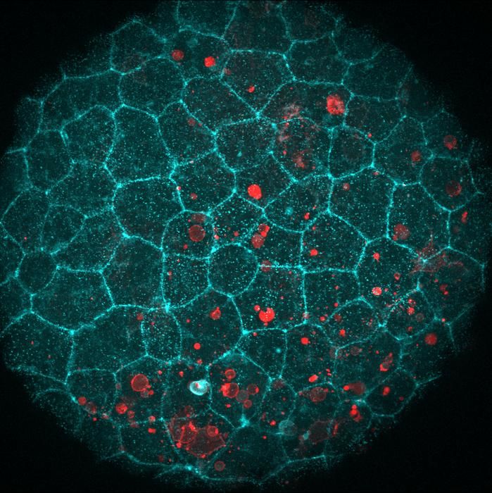
Featured image: Laura F. Bianchi/Centro de Regulación Genómica
Endnotes
- Biswas, S., et al. “Leiomodin 2 is a processive pointed-end elongator of actin filaments.” Nature Communications (published July 20, 2026). DOI: 10.1038/s41467-026-74809-z.
- Emory University. “Scientists solve mystery of how muscle cells grow and stay healthy.” News release via EurekAlert!, July 29, 2026. https://www.eurekalert.org/news-releases/1138119


Leave a Reply