
Nagoya Institute of Technology researchers showed that three amino acid changes reshape the electrical environment around a single light-catching molecule to separate red from green.
The ability to tell a ripe red fruit from the green leaves around it is something most people take for granted. It depends on a remarkably small piece of molecular machinery. Researchers at Nagoya Institute of Technology in Japan have now captured that machinery in detail for the first time, revealing how primate eyes distinguish red from green.
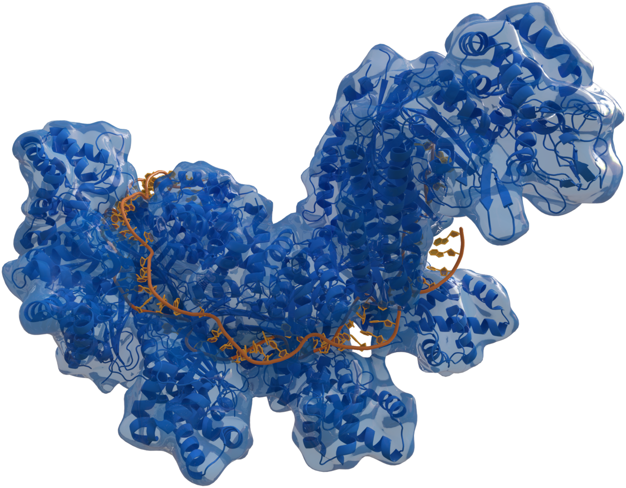
“This study reports the first three-dimensional structures of primate red- and green-sensitive cone visual pigments in their dark state,” said Dr. Kota Katayama, an associate professor of life science and applied chemistry. The work is published in Science.
One molecule, three colors
Human color vision relies on three types of cone cells in the retina, each most sensitive to a different range of wavelengths: short (blue), medium (green) and long (red). The brain compares their signals to produce the full range of colors we perceive.
The green and red cones have peak sensitivities around 530 and 560 nanometres respectively, close enough that their responses overlap heavily. It is precisely that small offset, about 30 nanometres, that lets the brain compare the two signals and distinguish reds, oranges, yellows and greens.
Strikingly, all three cone types use the same light-absorbing molecule, 11-cis-retinal, a derivative of vitamin A. When it absorbs a photon, it changes shape, triggering a cascade of signals that ends in a nerve impulse. What sets each cone’s color sensitivity is not the retinal itself but the protein wrapped around it, called an opsin.
The red and green opsins are especially close relatives. Their genes sit side by side on the X chromosome and are nearly identical, the product of a gene duplication in the ancestors of Old World monkeys, apes and humans that gave those primates three-color vision. That closeness has a cost. Because the genes are so similar, they can recombine unevenly, which is the main reason red-green color blindness is common, affecting about 8 percent of men of Northern European ancestry and far fewer women.
Many other mammals, including dogs and cats, have only two cone types and see the world with a more limited palette. Among New World monkeys, color vision varies even within a species, because many males inherit only one version of the X-linked opsin gene, while females who inherit two different versions can see in three colors.
Hard to pin down
Biologists have known for decades that small differences in the opsins’ amino acid sequences shift their color sensitivity. Studies in the 1980s and 1990s pinpointed a handful of positions that appeared to account for most of the difference between red and green pigments. But explaining exactly how those differences work has required seeing the proteins’ structures, and cone pigments are notoriously unstable and difficult to study.
Rhodopsin, the pigment in the rod cells responsible for dim-light vision, has been studied in structural detail since its first crystal structure was published in 2000. The cone pigments had remained out of reach.
Katayama and colleagues, including Massimo Olivucci of the University of Siena and Bowling Green State University, Hideaki Kato of the University of Tokyo and Hideki Kandori of Nagoya Institute of Technology, determined the structures of red and green cone pigments from the crab-eating macaque, Macaca fascicularis. The release notes that these pigments share about 98 percent of their amino acid sequence with their human counterparts.
Three small changes
The team compared the two structures and built detailed computer models of the pocket that holds the retinal. They focused on three positions where the pigments differ, then created mutant proteins carrying red-pigment residues at those positions and measured how the changes shifted light absorption.
The results showed that three amino acid substitutions, labelled A180S, F277Y and A285T, account for nearly all of the roughly 30-nanometre difference in peak light absorption between the red and green pigments. Those are the same three positions earlier genetic and spectroscopic studies had implicated, and the new structures show how they work.
Surprisingly, the substitutions do not substantially change the shape of the retinal. Instead, they change the electrical environment around it. The largest effect comes from a threonine residue whose hydroxyl group sits close to the chromophore, altering the distribution of charge that the light-absorbing molecule feels.
“Small sequence changes tune color by reshaping the electrostatic environment around the retinal chromophore,” Katayama said.
A hidden doorway
The structures also revealed something unexpected: a previously unknown opening in the cone pigments that faces the cell membrane and is absent in rhodopsin.
After a pigment absorbs light, its spent retinal must be replaced with a fresh molecule before it can respond again. Cone pigments regenerate much faster than rhodopsin, which helps cones work in bright, rapidly changing light. The team suggests the membrane-facing opening may help explain that speed by giving retinal an easier way in and out.
Katayama said that “distinct membrane-access pathways enable efficient retinal exchange and rapid visual pigment regeneration.”
From color blindness to drug design
The findings could help researchers understand how genetic variations alter color perception and contribute to color vision deficiencies and other visual disorders. “In the long term, this knowledge may help researchers understand how genetic variations alter color perception,” Katayama said.
There may be wider implications, too. Visual pigments belong to the family of G protein-coupled receptors, the largest family of cell-surface receptors in the human body and the target of roughly a third of approved drugs. The authors suggest the structural insights could inform drug discovery for photoreceptors and other receptors in the family.
The release does not report the number of samples or measurements, or state limitations of the work. The structures capture the pigments in their dark state, before they absorb light, and further work will be needed to follow how they change when they are activated.
Notes
- “Structural insights into spectral tuning and retinal exchange in cone visual pigments.” Science 392, no. 6805 (2026). Published 25 June 2026. DOI: 10.1126/science.adz3996.
- Nagoya Institute of Technology. “Scientists at Nagoya Institute of Technology uncover molecular basis of red–green color vision.” EurekAlert!, 27 July 2026. https://www.eurekalert.org/news-releases/1137598.
Featured image: Photo by Skitterphoto on Pexels.com



Leave a Reply