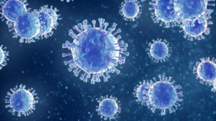
Indian Institute of Science researchers captured the encephalomyocarditis virus’s RNA gripping the host ribosome directly, exposing a possible drug target shared with poliovirus.
Viruses are the ultimate freeloaders. They carry instructions for making new viruses but none of the machinery needed to carry those instructions out. To replicate, they must commandeer the infected cell’s ribosomes, the molecular factories that build proteins, and persuade them to churn out viral proteins instead of the cell’s own.
Researchers at the Indian Institute of Science (IISc) in Bengaluru have now captured, in molecular detail, how one virus pulls off that takeover. Writing in eLife, the team used cryo-electron microscopy to show how the encephalomyocarditis virus, or EMCV, grabs hold of the host’s protein-making machinery.
A virus with a back door
EMCV is spread mainly by rodents and can infect a wide range of mammals, causing inflammation of the heart and brain, reproductive problems and neurological disorders. It belongs to the picornaviruses, the same family as poliovirus.
Normally, a cell’s ribosomes recognize the start of a messenger RNA by a chemical cap at its front end, then scan along until they find the instructions for a protein. Many viruses skip that process. Their RNA contains a folded region called an internal ribosome entry site, or IRES, that recruits ribosomes directly. Viruses often simultaneously shut down the cap-dependent translation of the host’s own genes, so the cell’s factories end up working almost exclusively for the virus.
IRES elements were first discovered in EMCV and poliovirus in the late 1980s. But the details of how they actually seize the ribosome have remained murky. “Nothing was known in terms of the details of how [the virus] captures the host ribosome other than the translational factors involved – and how exactly it does so biochemically,” said Tanweer Hussain, an associate professor in the Department of Developmental Biology and Genetics at IISc and the study’s corresponding author.
Fishing out a fragile complex
The first step in making viral protein is the formation of what is called a pre-initiation complex, in which the viral RNA, the small 40S subunit of the ribosome and several helper molecules come together. Capturing that complex intact is notoriously difficult.
Deepakash Das, a PhD student in Hussain’s lab and the study’s first author, designed a pulldown method to do it. Using a messenger RNA containing the EMCV IRES as bait, along with a specially designed bait protein, the team fished the complex out of rabbit reticulocyte lysate, an extract of immature red blood cells widely used to study protein synthesis. The catch included the 40S subunit, the initiator transfer RNA that begins every protein chain, and a helper complex called eIF2.
It did not always go smoothly. “Early purification attempts went well – something of a beginner’s luck – but many subsequent rounds failed to produce usable results,” Hussain said. The imaging was equally stubborn. “Cryo-EM grids frequently failed to yield enough particles for a reliable structure, but we persisted.”
A direct grip
Cryo-electron microscopy, whose development earned the 2017 Nobel Prize in Chemistry, freezes molecules in place and images thousands of them to reconstruct their three-dimensional shape. “CryoEM is the go-to technique that can actually give us a real-world picture of whatever is happening,” Das said.
The resulting structure showed that the EMCV IRES binds directly to both the 40S ribosomal subunit and the initiator transfer RNA, a mechanism the researchers say has not been seen in other viruses. Part of the viral RNA appears to mimic a section of the host’s own ribosomal RNA, a molecular disguise that may help it slot into place.
That mimicry also resembles a corresponding region in the poliovirus IRES, suggesting the two viruses, which belong to different branches of the picornavirus family, may share a common hijacking strategy. If so, a drug designed against one might work against both.
A target the host doesn’t need
The most promising aspect for drug developers is that the interface the IRES uses is not part of the host cell’s normal translation process. That raises the possibility of blocking the virus without disrupting the cell.
“We wanted to have a full picture of the IRES and ribosomal interactions so that we can design inhibitors targeting this region later,” Das said.
“We hope to selectively target only the viral translation pathway, leaving host translation unaffected,” Hussain said. “This is what we plan to test next, using mimics and candidate drug compounds.”
EMCV itself is more than a laboratory curiosity. It has caused deadly outbreaks of heart inflammation in pigs and has killed animals in zoos, from elephants to primates. Its close relative, poliovirus, is the target of one of the largest public health campaigns in history, and although wild poliovirus has been pushed back to a handful of countries, the eradication effort is not finished. The wider picornavirus family also includes the enteroviruses behind hand, foot and mouth disease and the rhinoviruses that cause many common colds, so insight into how these viruses commandeer ribosomes has broad relevance.
The work is a structural study conducted in a cell extract, not in infected animals, and the release does not report the resolution of the structures. Any drug candidates remain to be designed and tested. But by revealing exactly where and how the viral RNA grips the ribosome, the study turns a long-standing black box into a map, one that could guide antivirals against EMCV, and perhaps against its better-known relative, poliovirus, as well.
Notes
- Das, D., et al. “Structural insights into the recruitment of viral type 2 IRES to ribosomal preinitiation complex for protein synthesis.” eLife, 25 June 2026. DOI: 10.7554/eLife.107788.3.
- Indian Institute of Science. “How the encephalomyocarditis virus hijacks host protein machinery.” EurekAlert!, 23 July 2026. https://www.eurekalert.org/news-releases/1137280.



Leave a Reply