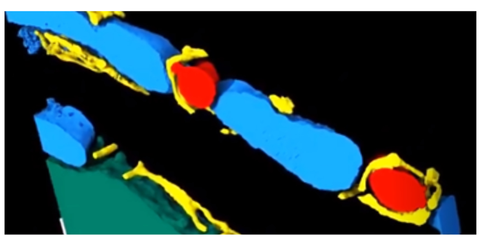
What happens below the cellular level when the heart contracts and relaxes has long been unexplored. Thanks to new ultra-high-resolution electron microscopy techniques, scientists can now watch the heart beating – almost at a molecular level. Researchers at the Medical Faculty of the University of Freiburg summarise the most important developments in cardiac electron microscopy and their significance for research in a recent publication, published in Nature Reviews Cardiology.

Insight at the nanometre scale is of great importance for the development of new therapies, for example for heart attacks or cardiac arrhythmias.
“With the high-resolution microscopy techniques developed by us and others worldwide, we gain fascinating insights into the dynamic ultrastructure of the heart,” says the study’s lead author Dr Eva Rog-Zielinska. She heads the 4D Imaging Section at the Institute of Experimental Cardiovascular Medicine (IEKM) of the University Heart Centre at the University Medical Centre Freiburg. “We can use this insight to analyse the three-dimensional structure of heart cells with unprecedented precision. Our images are made up of cubes – so-called voxels – with an edge length of one nanometre or less. For illustration: one nanometre is the distance a fingernail grows in one second,” Rog-Zielinska explains.
Watching the heart beat in super slow motion
A challenge is to link ultra-high resolution mapping of the heart to a moving target. “Thanks to recent advances in imaging, we now have a much better understanding of how muscle and connective tissue cells behave in the beating heart,” says co-author Prof Peter Kohl, Director of the IEKM, who is also the spokesperson of the German Collaborative Research Centre 1425 dedicated to exploring cardiac scarring.
Electron microscopy itself, but crucially also newly developed methods for the preparation and post-processing of corresponding samples, play a central role in the generation of molecular insight. “It is particularly exciting that we can record muscle cells like individual frames in a film – thanks to millisecond-precise high-pressure freezing. This allows us to watch the heart‘s molecular structures beating in super slow motion, as it were,” says Kohl.
Experiments, simulations and artificial intelligence intertwine
The microscopic images are evaluated at IEKM with the help of artificial intelligence, assisted by computer simulations to depict heart function and pathological changes as realistically as possible. “Newly gained insight allows us to gain a completely new understanding of cardiac activity and, based on this, to develop new therapeutic concepts. We are looking forward to a very exciting time in heart research,” says Kohl.
IMAGE CREDIT: Medical Center – University of Freiburg



Leave a Reply