Words matter. Images matter. The Scientific Inquirer needs your support. Help us pay our contributors for their hard work. Visit our Patreon page and discover ways that you can make a difference. http://bit.ly/2jjiagi
Cheng Chao-Min (Institute of Biomedical Engineering, National Tsing Hua University), Cheng Nai-Chen (Department of Surgery, National Taiwan University Hospital), and Wu Yuan-Kun (Department of Internal Medicine, National Taiwan University Hospital) have studied biofilm formation in chronic wounds. They set aside time to discuss their latest research with SCINQ.
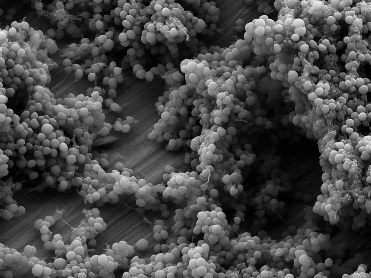
SCIENTIFIC INQUIRER: First for some background. What are biofilms? What role do they play in chronic wounds?
CHENG CHAO-MIN: Biofilms are the multi-species communities that aggregate together by a self-produced extracellular polymeric substance (EPS) matrix, and can attach on a wide variety of surfaces. The EPS matrix plays as a shield to protect the embedded microcolonies.
During the past decade, the clinical-based evidence has recognized that the bacterial infection is one of the important factors for both diagnosis and treatment of chronic wounds. It has indicated that 60% of the chronic wound has been characterized to mainly contain the biofilm. This kind of biofilm-based chronic wounds shows a better resistance to the conventional antibiotics, other antimicrobials, and in particular, the host defense systems.
SI: Why did you undertake a review of literature addressing biofilms in chronic wounds?
CC: The wound beds dwelt with biofilms exhibit the prolonged wound healing process (more than 30 days), and usually require more medical care associated with the medical resource. With the increasing prevalence of diabetes and other chronic diseases, this biofilm-complicated chronic wound issue has emerged as a global public health dilemma.
Microbiology researchers have built approaches to examine the planktonic state of microorganisms, and have reported the susceptibility to various antibiotics and antiseptics under planktonic conditions. However, the specific antibiotic therapy based on the susceptibility testing of planktonic microorganisms may result in the treatment failure because the biofilm-associated infection (occurs to the chronic wound) exhibits the increased resistance to antibiotics as mentioned previously.
SI: Can you describe your methodology?
CC: We have established a colorimetric-based approach with the characteristics of the simplicity, inexpensiveness and time-saving, allowing the healthcare persons to rapidly check whether the biofilm forms on the wound beds or not. Our goal is to develop an easy-to-handle staining procedure to detect whether the biofilm forms on the wound beds or not. The colorimetric-based result can be easily viewed as the positive or negative result (i.e., binary test to know whether the biofilm forms on the wound beds or not) through just checking the color change via the naked eyes (or combined with the smartphone APP).
SI: In the section discussing current diagnosis methodology you state, “there are no unequivocal gold-standard tests currently available for the diagnosis of biofilms in wounds.” How has this hampered addressing biofilms in a clinical setting?
CC: It is currently based on the patients’ disease histories, signs and symptoms, microscopic findings, and specific immune responses to suspect whether a particular wound has been inflicted with the biofilm. The current diagnostic approach highly relies on the experience-based evaluations determined by the clinicians. However, the diagnosis (to the biofilm on the wound beds) might be different among clinicians with the different levels of experience in terms of the wound infection management. This is the main reason for us to state that there is an emerging clinically relevant need to build a reliable approach to specifically detect the biofilm on the wound beds in a short period of time, i.e., in the near future, wish to become a routine diagnostic approach for the biofiom determination.
SI: From your research, which diagnostic methods are most effective? How do they fall short?
CC: Culture-based identification of microbial pathogens is most frequently, a well-established and standard method in clinical settings. However, the traditional culturing methodology only identifies 1% of the bacteria in chronic wounds, because the culture yield rate is affected by the state of the bacteria. Biofilms contain a large number of cells in stationary phase (a slow-growing or stationary phase in the bacterial growth cycle) due to microenvironmental stresses. These slow-growing bacteria are known to enter a viable but nonculturable (VBNC) state. Besides, culturability is further complicated by multispecies-type biofilms.
SI: Are there any lines of investigation you came across while writing the review that stands out as essential for moving the field forward?
CC: Wound blotting is a newly proposed methodology to provide a non-invasive and precise evaluation of wound beds. By applying a nitrocellulose membrane on wound beds, biomolecules, such as proteins and mucopolysaccharides, can be absorbed and immobilized for further analysis. In addition to providing qualitative assessment of the desired biomolecule, this method also allows spatial analysis of the target molecule across the entire wound bed, which may be heterogeneously distributed.
Nakagami and colleagues at University of Tokyo recently used the wound blotting methodology to detect biofilm formation on the wound beds of pressure injury. Nitrocellulose membranes were applied after sharp debridement, followed by evaluation for mucopolysaccharide content (an EPS component) with ruthenium red. This approach appears quite promising in terms of the biofilm detection for various wounds.
SI: Finally, what is next for you in terms of research?
CC: Based on the wound blotting technique, we would like to test various biomaterials as a film to absorb wound bed biomolecules. Moreover, we will optimize the staining procedure to achieve timely and precise biofilm detection.
IMAGE SOURCE: Creative Commons


Leave a Reply