A chain of immune reactions in the gut—driven by a key signaling protein and a surge of white blood cells from the bone marrow—may help explain why people with inflammatory bowel disease (IBD) have a higher risk of colorectal cancer, according to a preclinical study by Weill Cornell Medicine investigators. The findings point to new possibilities for diagnosis, monitoring and treatment.
The study began with a focus on TL1A, an inflammatory immune signaling protein known to be associated with IBD and colorectal cancer. Experimental drugs that block TL1A activity have shown great promise against IBD in clinical trials, but how the signaling protein promotes the disease and associated tumors has been unclear. The study, published in Immunity, demonstrates that in an animal model TL1A has much of its impact through immune cells in the gut called ILC3s. When these cells are activated by TL1A, they summon a surge of white blood cells called neutrophils from the bone marrow and reprogram them in ways that effectively promote tumor formation.
“These findings are important given the intense interest in the medical community to understand TL1A’s role in IBD and its potential role in associated colorectal cancers—for which we have had few strategies to mitigate the cancer risk,” said study senior author Dr. Randy Longman, director of the Jill Roberts Center for Inflammatory Bowel Disease at Weill Cornell Medicine and NewYork-Presbyterian/Weill Cornell Medical Center and an associate professor of medicine at Weill Cornell Medicine.

IBD, which includes Crohn’s disease and ulcerative colitis, is characterized by chronic gut inflammation. Between 2.4 and 3.1 million Americans have the condition, according to the U.S. Centers for Disease Control and Prevention. IBD raises the risk of other autoimmune and inflammatory conditions and greatly increases the risk of colorectal cancer, which tends to occur at younger ages and with worse outcomes in patients with the condition.
In the study, Dr. Longman’s team discovered that TL1A, which is produced mostly by other immune cells in the IBD gut, works to stoke tumor growth principally through gut-resident ILC3 cells. When activated by TL1A, these cells secrete a blood cell growth factor called granulocyte-macrophage colony-stimulating factor (GM-CSF). This in turn triggers a process called “emergency granulopoiesis”—a burst of new neutrophil production in bone marrow—followed by the influx of the neutrophils to the gut. In mouse models of gut cancer, adding such neutrophils was enough to promote tumor development.
Neutrophils can promote colorectal tumors by secreting highly reactive molecules that can damage DNA in gut-lining cells. However, the team found that the gut ILC3s also induce a distinctive pattern of gene activity in the neutrophils including increased expressions of genes known to promote tumor initiation and growth. The researchers observed a similar gene activity pattern in samples of colitis-affected gut tissue from patients with IBD, and this tumor-promoting signature was less evident in patients who took an experimental TL1A-blocking treatment.
The results suggest that not only TL1A but also ILC3s, GM-CSF, and ILC3-summoned neutrophils could be targets in future strategies to treat IBD and prevent associated colorectal tumors.
“I think it will be exciting for clinicians in the IBD field to know that there is a systemic process at work here, involving both the gut and the bone marrow, with the potential to drive precision medicine in IBD,” said study first author Dr. Sílvia Pires, an instructor in medicine and member of the Longman Laboratory.
The team now is following up with further studies of this cell communication pathway in the context of gut inflammation, including the potential early role of occasional GM-CSF exposure in sensitizing marrow cells so that IBD becomes increasingly likely.
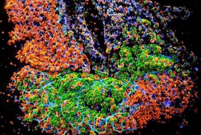
IMAGE CREDIT: Longman Lab.


Leave a Reply