Instinctive fear responses—like fleeing from a sudden shadow overhead—are crucial for survival. Yet, animals (including humans) often learn to suppress these reactions when they realize a stimulus is harmless. A new study by neuroscientists at the Sainsbury Wellcome Centre at University College London uncovers how this learning process unfolds in the brain, revealing that the visual cortex plays a pivotal role during learning but not in storing the memory. Instead, the memory is maintained in the ventrolateral geniculate nucleus (vLGN), a subcortical structure.
In this Q&A, Prof. Sara Medros, co-lead author of the study, delves into their experiments with mice, the significance of their findings, and potential implications for understanding and treating anxiety disorders and PTSD in humans.
What inspired your team to explore how animals learn to suppress instinctive fear?
Our interest began with earlier work from our lab showing that the ventrolateral geniculate nucleus (vLGN) plays a key role in regulating instinctive fear responses—hard-wired defensive behaviors such as escape or freezing that occur without any prior experience. For example, when an animal detects a potential approaching predator such as a bird from above, it instinctively escapes toward shelter, even without identifying the exact nature of the threat.
What made this especially intriguing is that animals can naturally learn to suppress these responses when the stimulus turns out to be harmless. This is a very simple form of natural learning, driven purely by experience, and doesn’t require any external training or reinforcement. Because this type of behaviour is both ecologically relevant and easy to observe, we were curious to understand how the brain supports this kind of adaptation.
Could you explain your experiments with mice and visual threats in simple terms?
To simulate a naturalistic visual threat, we projected an expanding black disk onto a screen above the animal, mimicking the approach of a predator from above. This looming stimulus is known to evoke instinctive escape behaviour in mice. In our setup, the mice were placed in an arena with a shelter—a small enclosed area they instinctively recognise as a safe space.
Initially, when the disk appeared, the mice responded by escaping to the shelter. However, after repeated exposures where no actual harm followed, they began to adapt. The same stimulus no longer triggered an escape response, and instead, the animals explored the arena more freely. This shift allowed us to study how the brain learns that a previously threatening stimulus no longer predicts danger, and which neural circuits are involved in this process.
Why is it important for animals—including humans—to override instinctive fear responses?
Instinctive fear responses are critical for survival, as they enable rapid reactions to potential threats. But continuing to respond to stimuli that turn out to be harmless can be costly. For animals, that could mean expending unnecessary energy or missing opportunities for foraging, mating, or exploring.
In modern humans, the types of threatening experiences we face are often different. We may no longer need to worry about predators, but we encounter other types of stressful or challenging situations in daily life—like public speaking, job interviews, or unfamiliar social settings. At first, these can provoke strong emotional and physiological responses, but with experience, we often learn that they are not harmful. For example, children may initially fear fireworks due to their loud and unpredictable nature, but eventually learn they are harmless and even enjoyable. The ability to override instinctive fear is therefore essential for flexible, adaptive behaviour.

Your study found that specific areas of the visual cortex help animals learn not to fear previously threatening situations. Why is this significant?
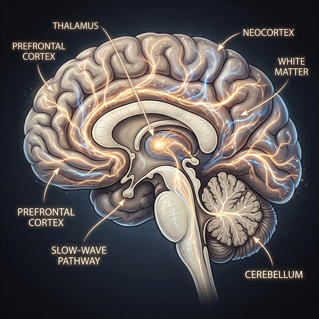
We found that the visual association cortex—higher visual areas in the brain—plays a crucial role in helping animals learn to suppress instinctive fear. This area sends projections to the vLGN and appears to instruct the learning process that allows animals to adapt their defensive responses.
What’s striking is that this cortical input is essential during learning but not afterward. Once the animal has learned that the stimulus is safe, the visual cortex is no longer required to maintain that behavioural change. This suggests that the cortex acts like a teacher—it helps establish the new response but doesn’t store it. Instead, the memory is stored in downstream subcortical circuits. This challenges the traditional view that memory and learning-related plasticity primarily occur in cortical areas and highlights a previously underappreciated role for subcortical structures like the vLGN.
After mice learn not to fear something, the visual cortex isn’t needed anymore. Where is this new “memory” stored in the brain?
In our study, the memory of the learned overwriting of fear responses appears to be stored in the vLGN. With experience, inhibitory connections onto the vLGN neurons that can suppress the animal’s escape response are weakened, and this change in connections persists over time. This synaptic plasticity change is mediated by endocannabinoid signalling, which drives a form of inhibitory long-term depression. As a result, the response of a subset of vLGN neurons to the visual threat stimulus is increased, allowing them to exert stronger suppression on downstream fear circuits and reduce escape behaviour.
You highlighted a brain region called the vLGN—how does this area control fear reactions?
The vLGN functions as an inhibitory hub that regulates the expression of instinctive fear responses. It receives input from the visual cortex and other areas and sends inhibitory projections to brain regions that drive escape behaviour. By suppressing the activity of these downstream circuits, the vLGN can effectively control whether or not a defensive response is expressed.
We found that with learning, endocannabinoid signalling in the vLGN mediates a form of inhibitory long-term depression, which reduces inhibition onto a subset of vLGN neurons. This makes them more active in response to the visual stimulus, increasing their ability to inhibit the escape circuit. In this way, the vLGN becomes a key site for modulating whether instinctive fear is expressed or suppressed based on prior experience.
Could your findings help us better understand or treat anxiety disorders and PTSD in humans?
Yes. The neural circuits we studied in mice, including the pathway from the visual association cortex to the vLGN, are also present in the human brain. In conditions such as anxiety disorders or PTSD, individuals often continue to respond to harmless stimuli as if they are dangerous. This may reflect a failure in the brain’s ability to suppress fear after learning that a stimulus is no longer threatening.
Our findings suggest that the vLGN—or its human equivalent, referred to as the pregeniculate nucleus—could be involved in this failure of suppression. If this brain region is not properly engaged, fear responses may remain exaggerated and maladaptive. This opens up potential avenues for treatment, such as targeting endocannabinoid signalling or using neuromodulation techniques like deep brain stimulation or focused ultrasound. These approaches could help restore the brain’s ability to regulate fear and improve outcomes for individuals affected by these disorders.
What’s the next step—what new questions does your research raise?
We’re now interested in exploring whether the same circuits involved in suppressing escape to visual threats also play a role in more general forms of fear regulation. For example, are they involved in situations of chronic stress or in helping animals cope with uncertain or changing environments? We are also interested in exploring whether the same circuits contribute to broader aspects of behavioural regulation, such as courage or adaptive decision-making in aversive situations.
Another direction is to understand whether these circuits are affected in models of anxiety or stress-related disorders, and whether manipulating them can enhance resilience. More broadly, we’re asking whether similar learning processes and circuit mechanisms apply to other types of sensory input or behavioural contexts. Our goal is to build a deeper understanding of how the brain adapts to experience and how this flexibility might be disrupted in disease.
Sign up for the Daily Dose Newsletter and get the morning’s best science news from around the web delivered straight to your inbox? It’s easy like Sunday morning.


Leave a Reply