
Ushikuvirus, isolated from a freshwater pond, joins a growing family of giant viruses that may have played a role in the evolution of eukaryotic cells
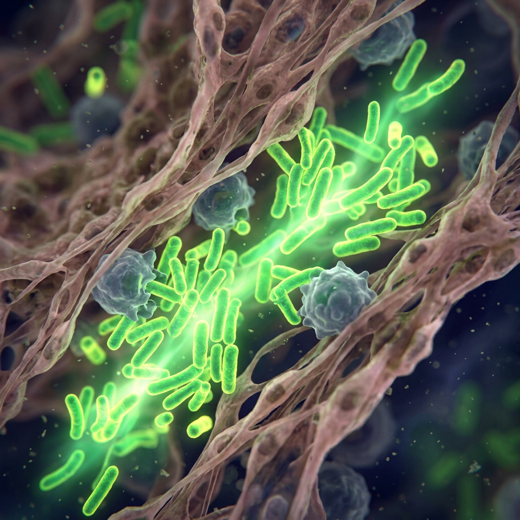
More than two decades after scientists first discovered that viruses could grow to enormous sizes, researchers continue to unearth new species that challenge our understanding of these enigmatic entities. The latest addition to this expanding catalog is ushikuvirus, a giant virus isolated from a freshwater pond in Japan that exhibits unusual features never before seen in its close relatives.
Named after Lake Ushiku in Ibaraki Prefecture, where it was collected, ushikuvirus represents the first giant virus isolated from Japanese aquatic environments that infects Vermamoeba vermiformis, a type of single-celled amoeba. The discovery, published in the Journal of Virology, adds to a family of viruses that scientists believe may hold important clues about how complex cells with nuclei—the hallmark of eukaryotic life—first emerged on Earth.
“Giant viruses can be said to be a treasure trove whose world has yet to be fully understood,” said Professor Masaharu Takemura of Tokyo University of Science, who led the research team. “One of the future possibilities of this research is to provide humanity with a new view that connects the world of living organisms with the world of viruses.”
The discovery of giant viruses fundamentally reshaped virology beginning in 2003, when French researchers identified Acanthamoeba polyphaga mimivirus in a water cooling tower. With a genome of 1.2 million base pairs and particle size of 400 nanometers, mimivirus shattered the long-held assumption that viruses were inherently tiny. Since then, more than ten new families of giant viruses have been discovered, revealing extraordinary diversity in size, shape, and genetic content.
What makes these viruses particularly fascinating to evolutionary biologists is that many encode genes once thought exclusive to cellular life. Some carry genes for making proteins, synthesizing DNA, and even packaging their genetic material into nucleosome-like structures using histone proteins—the same proteins that organize DNA within the nuclei of plants, animals, and fungi.

A Controversial Hypothesis
Takemura has long been a proponent of a provocative idea known as viral eukaryogenesis. In 2001, he and Australian researcher Philip Bell independently proposed that the nucleus of eukaryotic cells may have originated from a large DNA virus that infected an ancestral archaeal cell. Rather than killing its host, the virus established a long-term presence inside the cytoplasm, eventually evolving into the membrane-bound compartment we now recognize as the cell nucleus.
While this hypothesis remains contentious among scientists, the discovery of giant viruses that replicate within host cell nuclei and encode eukaryote-like genes has provided tantalizing circumstantial evidence. Members of the Mamonoviridae family, particularly the medusaviruses discovered since 2019, encode full sets of histone proteins and depend heavily on their hosts’ nuclear machinery for replication.
A recent structural study published in Nature Communications revealed that medusavirus histones can form nucleosome-like structures similar to those found in eukaryotic cells, though with notable differences suggesting adaptation to the virus’s unique lifecycle. These findings add weight to the possibility that gene exchange between ancient viruses and their hosts played a role in shaping modern eukaryotic cells.
A Distinct Relative
Ushikuvirus is closely related to clandestinovirus, discovered in French wastewater in 2021, and more distantly to the medusaviruses. Its genome spans at least 666,605 base pairs and contains 784 genes, making it larger than its closest relatives. Like other members of this viral group, ushikuvirus encodes a complete set of histone proteins, though its H2A and H2B genes are fused together—a configuration also seen in clandestinovirus and marseilleviruses.
But ushikuvirus displays several features that distinguish it from its relatives. Using cryo-electron microscopy, the researchers revealed that the virus’s icosahedral capsid is decorated with multiple spike-like “cap” structures, some bearing filamentous extensions not observed in medusaviruses. The capsid measures approximately 250 nanometers in diameter, excluding the spikes.
Perhaps most striking is how ushikuvirus affects its host. Unlike most giant viruses that cause infected cells to shrink, ushikuvirus causes vermamoeba cells to enlarge dramatically—on average doubling in size, with some individual cells growing to seven times their original dimensions. The infection cycle is also notably longer than that of related viruses, spanning approximately 60 hours or more.
“The discovery of a new Mamonoviridae-related virus, ‘ushikuvirus,’ which has a different host, is expected to increase knowledge and stimulate discussion regarding the evolution and phylogeny of the Mamonoviridae family,” Takemura explained. “As a result, it is believed that we will be able to get closer to the mysteries of the evolution of eukaryotic organisms and the mysteries of giant viruses.”

Breaking Down Barriers
Another unusual characteristic sets ushikuvirus apart from its closest relatives. While medusaviruses and clandestinovirus replicate within the intact host cell nucleus, ushikuvirus destroys the nuclear membrane as infection progresses. This behavior has been observed in pandoraviruses—giant viruses with genomes exceeding 2 million base pairs—but is unprecedented in the Mamonoviridae-related group.
The virus also forms specialized structures called viral factories within the host cytoplasm, where new virus particles are assembled. These factories appear as dense globular regions visible under electron microscopy, similar to those formed by mimiviruses. After assembly, progeny virions are released from infected cells primarily through exocytosis rather than cell lysis, allowing the host to survive longer while gradually releasing new viruses.
The researchers suggest that ushikuvirus’s unique surface structures—including fibrous projections that may contain carbohydrates—could explain its longer infection cycle and unusual effects on host cells. Studies of mimiviruses have shown that surface fibrils play important roles in viral attachment to host cells, and viruses lacking these structures replicate faster and induce cell death more quickly.

An Expanding Field
Giant viruses have become one of the most dynamic areas of virology research. A comprehensive review published earlier this year in npj Viruses noted that the past two decades have seen the discovery of more than ten new families of protist-infecting DNA viruses, with extraordinary diversity in virion shape, size, gene content, and replication strategies. These discoveries have forced scientists to reconsider traditional definitions of viruses and their place in the tree of life.
Several giant viruses that infect vermamoeba have now been isolated, including faustovirus, fadolivirus, orpheovirus, and kaumoebavirus. Some, like tupanvirus discovered in Brazil, can infect both vermamoeba and acanthamoeba. The diversity of these viruses and their hosts suggests that giant viruses are ubiquitous in aquatic and soil environments worldwide.
Beyond their evolutionary significance, understanding how giant viruses interact with amoebae may have practical implications. Certain Acanthamoeba species can cause serious human infections, including amoebic encephalitis. Learning how giant viruses destroy their amoeba hosts could potentially inform new strategies for treating such infections.
For now, ushikuvirus joins a growing roster of giant viruses that continue to surprise scientists with their complexity and diversity. Each new discovery provides another piece of a puzzle that may ultimately help explain one of biology’s deepest mysteries: how the first complex cells with nuclei emerged from simpler ancestors billions of years ago.
Sources
1. Bae J, Hatori N, Burton-Smith RN, Murata K, Takemura M. A newly isolated giant virus, ushikuvirus, is closely related to clandestinovirus and shows a unique capsid surface structure and host cell interactions. Journal of Virology. 2025;99(12). doi:10.1128/jvi.01206-25
2. Tokyo University of Science. Ushikuvirus: A newly discovered giant virus may offer clues to the origin of life. EurekAlert! January 7, 2026.
3. Bosmon T, Abergel C, Claverie JM. 20 years of research on giant viruses. npj Viruses. 2025;3:9. doi:10.1038/s44298-025-00093-1
4. Toner CM, Hoitsma NM, Weerawarana S, Luger K. Characterization of Medusavirus encoded histones reveals nucleosome-like structures and a unique linker histone. Nature Communications. 2024;15:9138. doi:10.1038/s41467-024-53364-5
5. Takemura M. Medusavirus ancestor in a proto-eukaryotic cell: updating the hypothesis for the viral origin of the nucleus. Frontiers in Microbiology. 2020;11:571831. doi:10.3389/fmicb.2020.571831
6. Yoshikawa G, Blanc-Mathieu R, Song C, et al. Medusavirus, a novel large DNA virus discovered from hot spring water. Journal of Virology. 2019;93:e02130-18. doi:10.1128/JVI.02130-18
7. Rolland C, Andreani J, Sahmi-Bounsiar D, Krupovic M, La Scola B, Levasseur A. Clandestinovirus: a giant virus with chromatin proteins and a potential to manipulate the cell cycle of its host Vermamoeba vermiformis. Frontiers in Microbiology. 2021;12:715608. doi:10.3389/fmicb.2021.715608
8. Gaïa M, Forterre P. From Mimivirus to mirusvirus: the quest for hidden giants. Viruses. 2023;15:1758. doi:10.3390/v15081758
9. La Scola B, Audic S, Robert C, et al. A giant virus in amoebae. Science. 2003;299:2033. doi:10.1126/science.1081867


Leave a Reply