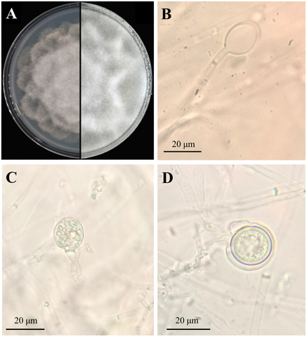

Phytophthora cinnamomi is a devastating plant pathogen, responsible for root rot in hundreds of plant species. Classified as an oomycete (a fungus-like microorganism), its life cycle displays a fascinating variety of forms—each captured in this composite microscopy image.
Panel A shows the seven-day-old colony grown on selective PARP medium. From this mat emerge reproduction structures: panel B reveals a sporangium, a sac-like structure that releases motile zoospores—tiny swimming spores—when submerged. These flagellated zoospores swim through soil water, seeking host roots to infect.
Panel C shows a gametangium, the sexual reproductive organ. In contrast to sporangia, gametangia form when resources are low or environmental signals trigger sexual reproduction. Fertilization produces oospores—shown in panel D—a durable thick-walled stage capable of surviving harsh soil conditions for years.
Once favorable cues return, oospores germinate to release zoospores or germ tubes, restarting infection. This dual lifestyle—sexual oospores for long-term survival and asexual zoospores for rapid spread—makes P. cinnamomi highly adaptive and persistent.
So why does this matter? As a pathogen, P. cinnamomi infects roots, cutting off water and nutrient transport. In forests, it causes dieback in native vegetation; in agriculture, it reduces crop yields and increases management costs. Recognizing the morphology in this image helps plant pathologists detect infection stages, predict outbreaks, and deploy treatment—such as fungicides or resistant plantings.
Technically, this image derives from open-access transmission and light microscopy described in a peer-reviewed study under CC BY license. The high resolution (1,772 × 1,944 px) allows scientists to distinguish subtle features like gametangial stalks and oospore wall thickness—key diagnostic traits. Highlighting these stages shows how microscopy remains essential in plant biosecurity, bridging molecular biology, ecology, and agronomy to manage a pathogen that threatens ecosystems globally.
IMAGE CREDIT: Xiaoqing Tong, Jiayi Wu, Li Mei & Yongjun Wang


Leave a Reply