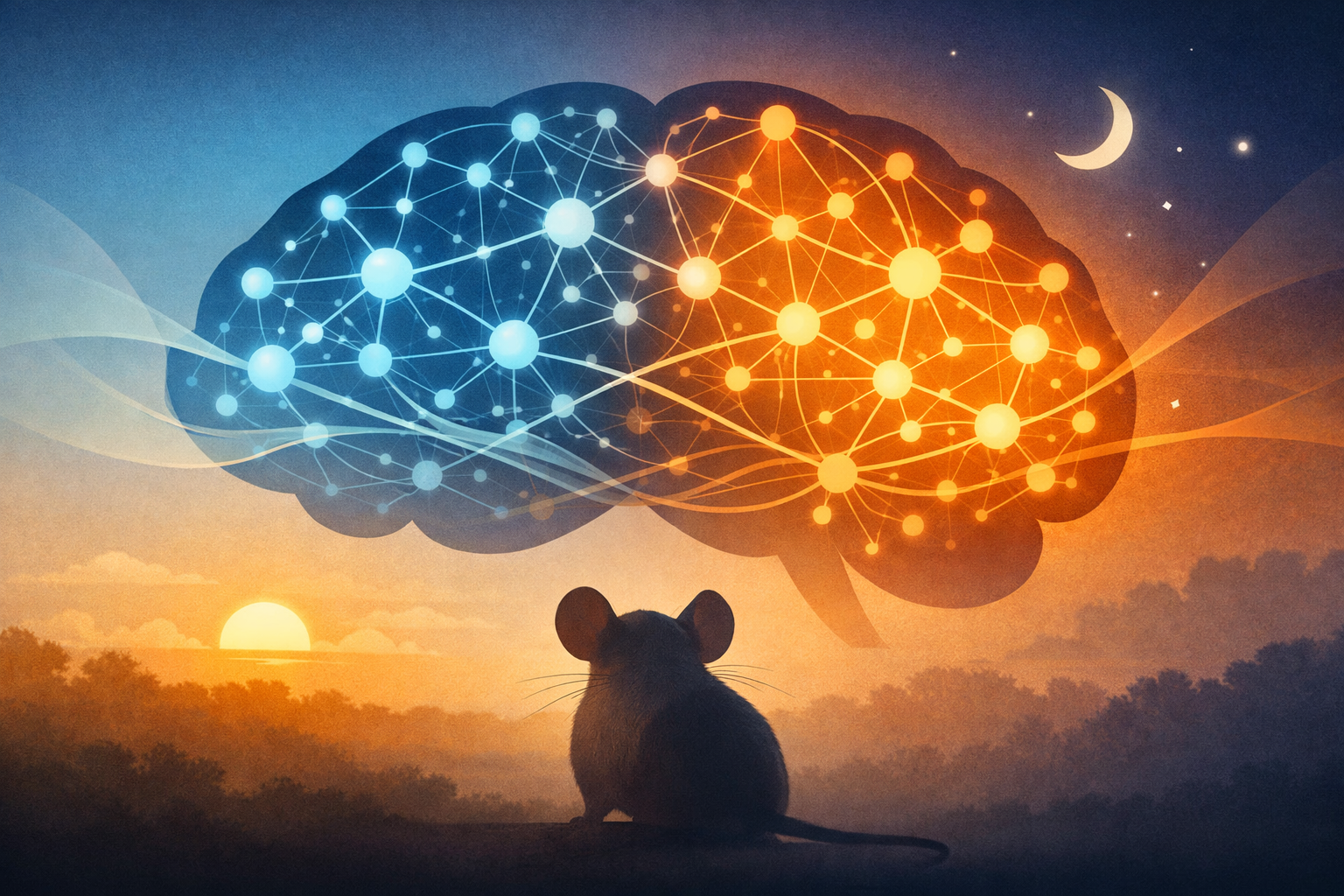
The mouse brain contains roughly 80 million neurons, yet until now, scientists have lacked the tools to observe which of these cells are active across the entire brain at different times of day. A groundbreaking international collaboration has changed that, developing an experimental and computational framework that reveals how neural activity dramatically reorganizes itself from morning to night—findings that could eventually help us understand human fatigue, sleep disorders, and psychiatric conditions.
Published in PLOS Biology, the study brought together researchers from the University of Michigan, the University of Zurich, and Japan’s RIKEN Center for Biosystems and Dynamics Research. Their work captures something remarkable: the brain doesn’t simply become more or less active as the day progresses. Instead, it fundamentally reorganizes which regions and networks take charge.
“We undertook this difficult study to understand fatigue,” said Daniel Forger, professor of mathematics at the University of Michigan and the study’s senior author. “We’re seeing profound changes in the brain over the course of the day as we stay awake and they seem to be corrected as we go to sleep.”
The research team employed an ingenious approach. They used a genetic tagging system called TRAP2, which causes neurons to glow when they become active. By administering a drug at specific times—the beginning and end of both the rest period and the active period in mice—they could permanently mark which neurons were firing during each four-hour window. After allowing time for the fluorescent markers to develop, the researchers used advanced tissue-clearing techniques and light-sheet microscopy to create stunning three-dimensional maps of active neurons across entire brains.

A City’s Changing Traffic Patterns
What they discovered challenges simple notions of the brain as a static organ that merely dims and brightens. As mice began their active period (nighttime for these nocturnal creatures), neural hubs were concentrated in subcortical regions deep within the brain, including the posterior hypothalamic nucleus—an area known for regulating arousal and wakefulness. But as the night wore on, activity shifted dramatically toward the cortex, the brain’s outer layer responsible for higher cognitive functions.
“The brain doesn’t just change how active it is throughout the day or during a specific behavior,” explained Konstantinos Kompotis, a senior scientist at the University of Zurich’s Human Sleep Psychopharmacology Laboratory and study co-author. “It actually reorganizes which networks or communicating regions are in charge, much like a city’s roads serve different traffic networks at different times.”
The researchers found that by the end of the active period, the brain’s default mode network—a collection of regions that become active during rest and introspection—had assumed a more central position. Five of the top fifteen neural hubs during this pre-sleep window belonged to this network, compared to only two at the beginning of wakefulness. This shift may reflect the brain’s preparation for sleep and the consolidation of experiences from the day.

Layer by Layer
The study also revealed striking changes within the cortex itself. The cerebral cortex is organized into distinct layers, each with different types of neurons and connections. The researchers observed that layer 5 neurons—known for their role in communication between different cortical regions and for initiating the slow-wave activity characteristic of deep sleep—became significantly more active toward the end of the wake period.
This finding aligns with previous research showing that layer 5 neurons play crucial roles in both cortico-cortical communication and sleep regulation. The increased activity of these neurons may represent the brain’s mounting sleep pressure—the growing need for rest that accumulates during wakefulness.
Perhaps equally significant was the discovery of shifting balance between excitatory and inhibitory neurons. Throughout the brain, and particularly in the thalamus—a critical relay station for sensory information—the ratio of active excitatory neurons to inhibitory neurons changed across the day. At the end of the active period, inhibitory neurons became more prominent, which may relate to the increased demand for sleep-related processes as the day draws to a close.

Building on Decades of Brain Mapping
This research builds upon years of painstaking work to map the brain’s physical wiring. The Allen Brain Atlas, developed over the past decade, has provided increasingly detailed maps of structural connectivity between brain regions. More recent efforts, including the complete reconstruction of a block of the visual cortex with all its neuronal connections, have revealed the brain’s wiring diagram in unprecedented detail.
“We know from studies over the last 20 or 30 years, how to decipher how one aspect—a gene or a type of neuron, for instance—can contribute to behavior,” Kompotis noted. “But we also know that whatever governs our behavior, it’s not just one gene or one neuron or one structure within the brain. It’s everything and how it connects and interacts at a given time.”
But knowing how the brain is wired is only part of the story. Neural connections can vary in strength, and individual neurons can be more or less active depending on circumstances. The new study introduces the concept of “active connectivity”—a measure that combines structural wiring with actual neuronal activity to reveal which pathways are genuinely in use at any given moment.

From Mice to Humans
While the genetic tagging techniques used in this study cannot be applied to humans, the computational methods are transferable. Guanhua Sun, who worked on the project as a doctoral student at Michigan and is now at New York University, emphasized that the mathematical approaches could be adapted for human brain imaging data.
“The way we detect human brain activity is more coarse-grained than what we see in our study,” Sun explained. “But the method we introduced in this paper can be modified in a way that applies to that human data. You could also adapt it for other animal models, for example, that are being used to study Alzheimer’s and Parkinson’s. I would say it’s quite transferable.”
The potential applications are significant. Forger envisions developing objective “signatures” of fatigue that could help ensure people in high-stakes roles—pilots, surgeons, truck drivers—are adequately rested before performing critical tasks.
“We’re actually terrible judges of our own fatigue. It’s based on our subjective tiredness,” Forger said. “Our hope is that we can develop ‘signatures’ that will tell us if people are particularly fatigued, and whether they can do their jobs safely.”
The research also hints at connections to mental health. “This study doesn’t touch on that,” Forger acknowledged. “But I do think the activity we saw in different regions is going to be important for understanding certain psychiatric disorders.”
Kompotis has already begun collaborating with industrial partners to use the team’s techniques for evaluating how drugs and therapeutic candidates affect brain activity—a potentially powerful tool for pharmaceutical development.
A Collaborative Achievement
The study was supported by the Human Frontier Science Program, an organization dedicated to fostering international scientific collaboration, along with funding from the U.S. National Science Foundation and the U.S. Army Research Office. The researchers have made their data and computational tools freely available, enabling other scientists to build upon their work.
The team dedicated their paper to Steven Brown, a professor at the University of Zurich who was a core member of the collaboration before his death in a plane crash during the project.
“Steve was a perfect collaborator,” Forger recalled. “We learned how important one person can be in scientific research, be it in brainstorming or in bridging ideas and concepts,” Kompotis added. “Steve was a core element of this collaboration. It is yet another reason for us to be very proud of this story.”
As neuroscience continues to generate vast quantities of multimodal data—from gene expression to neural connectivity to real-time activity—studies like this one demonstrate the power of integrating different data types to understand the brain’s dynamic organization. The framework developed here captures only the most active one percent of the brain’s neurons, yet even this glimpse reveals a remarkably plastic organ that continuously reorganizes itself to meet the demands of waking life and the restorative needs of sleep.
Sources and References
1. Sun G, Mano T, Shi S, et al. A framework to determine active neurons and networks within the mouse brain reveals how brain activity changes over the course of the day. PLOS Biology. 2025;23(11):e3003472. doi:10.1371/journal.pbio.3003472
2. University of Michigan News Release: “How brain activity changes throughout the day.” December 8, 2025. EurekAlert.
3. Oh SW, Harris JA, Ng L, et al. A mesoscale connectome of the mouse brain. Nature. 2014;508(7495):207-14.
4. MICrONS Consortium. Functional connectomics spanning multiple areas of mouse visual cortex. Nature. 2025;640(8058):435-47.
5. Whitesell JD, Liska A, Coletta L, et al. Regional, layer, and cell-type-specific connectivity of the mouse default mode network. Neuron. 2021;109(3):545-559.
6. Kim EJ, Juavinett AL, Kyubwa EM, et al. Three types of cortical layer 5 neurons that differ in brain-wide connectivity and function. Neuron. 2015;88(6):1253-67.
7. Krone LB, Yamagata T, Blanco-Duque C, et al. A role for the cortex in sleep-wake regulation. Nature Neuroscience. 2021;24(9):1210-5.
8. Zhang M, Pan X, Jung W, et al. Molecularly defined and spatially resolved cell atlas of the whole mouse brain. Nature. 2023;624(7991):343-54.


Leave a Reply