When neuroscientists hunt for early signals of cognitive aging, they tend to chase the big, abstract measures—memory scores, fluid intelligence, executive function. But in a sweeping new analysis of nearly 28,000 adults from the UK Biobank, it is reaction time—simple, blunt, and deeply embodied—that emerges as the clearest indicator of how the aging brain reorganizes itself.
The finding goes deeper than the familiar observation that people slow down as they age. Reaction time, the researchers found, tracks tightly with an underlying architectural shift: a negotiation between structural decline and functional rewiring that unfolds unevenly across the brain. As the study’s authors put it, their work “unveils diverse joint function–structure changes, providing strong evidence for understanding distinct cognitive deteriorations during aging.”
What makes this study significant isn’t any single headline result. It’s a set of findings that, taken together, challenge the notion of brain aging as a uniform process. Aging, the data suggest, is not one trajectory but many—sometimes synergistic, sometimes contradictory, and sometimes lateralized in surprising ways.

The Patchwork Brain
One of the study’s most consequential insights is that brain aging doesn’t follow a single script. When researchers examined brain structure and function together, they identified two dominant patterns of change.
In some regions, gray matter volume and functional connectivity decline in lockstep. This is the intuitive story of neurodegeneration: less tissue, weaker communication, poorer performance. The study found that “synergistic joint changes (i.e., 9 of 15 changes) were predominant, involving both FNC and GMV decreased primarily in the cerebellum, frontal pole, paracingulate gyrus, and precuneus cortex.” These are regions long associated with motor coordination and cognitive control, and their parallel decline reinforces decades of research linking cortical thinning to slower, less reliable information processing.
Elsewhere, however, something more paradoxical emerges. Gray matter volume declines while functional connectivity increases. This contradictory pattern suggests compensation—as structural resources diminish, remaining networks appear to communicate more intensely to maintain performance. The researchers observed these “contradictory changes with increased FNC magnitude but decreased GMV in the occipital pole, lateral occipital cortex, and frontal pole, acting as a compensatory mechanism as one ages to preserve visual acuity, cognitive ability, and behavioral modulation.”
This aligns with the compensation-related utilization of neural circuits hypothesis (CRUNCH), which proposes that older brains recruit additional network activity to offset efficiency losses. But crucially, these two patterns coexist within the same individuals. The aging brain isn’t globally compensatory or globally degenerative—it’s patchwork. The same brain can show quiet collapse in one circuit and energetic over-engagement in another.

THE BOTTOM LINE

Your reaction time may be an early warning system for brain health.

- Simple tests catch problems early. Reaction time tracks brain changes more closely than memory or IQ tests—a quick, cheap way to spot decline before it's obvious.

- Your brain doesn't age all at once. Some areas lose structure and function together; others compensate by working harder. The pattern varies by person.

- Compensation has limits. When brain regions work harder to offset decline, it helps—but only temporarily, and at a cost.

- The cerebellum is key. Once dismissed as just a "motor" region, it's now central to understanding cognitive aging and reaction speed.

- Future potential. Since reaction time is easy to measure digitally, phones and wearables could one day monitor brain health like they track heart rate.
Why Reaction Time Matters
Why does reaction time emerge as the most sensitive marker? Because it sits at the intersection of perception, decision-making, and motor execution. Unlike memory recall or problem-solving tasks, reaction time offers little room for strategy. It reflects how quickly the brain can coordinate multiple systems under minimal cognitive cover.
The study found that reaction time correlates most strongly with joint changes in both structure and function—particularly in circuits linking the cerebellum and frontal midline regions. “The joint change most strongly associated with RT showed concurrent decreases in both FNC and GMV between the right crus I of the cerebellum (CB domain) and the right paracingulate gyrus (CC domain),” the researchers note. “This result indicates that age-related declines in both structural and functional integrity in these regions may directly contribute to slower cognitive reaction speed.”
The cerebellum—long miscast as a purely motor structure—is now recognized as a key player in timing, prediction, and cognitive coordination. Recent consensus papers describe it as building “internal models” that facilitate automatic behaviors through interactions with the cerebral cortex, basal ganglia, and spinal cord. Some evidence even suggests that cerebellar volume independently predicts scores on intelligence tests, and morphological research has found it often as good as—or better than—the prefrontal cortex at predicting cognitive performance.
This makes the cerebellum’s coupling with frontal control regions an ideal substrate for reaction-time effects. What’s compelling isn’t just the correlation itself but its selectivity. Fluid intelligence, often treated as the gold standard of cognitive aging measures, showed weaker associations with these joint brain changes. Reaction time, by contrast, tracked them closely.
This finding supports a growing body of work suggesting that processing speed decline is among the earliest and most pervasive features of aging, cascading into higher-order deficits later. As the study notes, “RT and NM exhibit stronger associations with joint changes compared to FI… These results are consistent with prior knowledge that FI tends to be more resilient to aging effects, while RT and memory-related functions are more vulnerable.”

Compensation Has Limits
The idea of compensatory hyperconnectivity is appealing—it suggests resilience, the brain fighting back against entropy. But the study is careful to show that compensation is neither universal nor free.
Regions showing increased connectivity alongside structural loss aren’t necessarily “healthier.” In some cases, heightened connectivity may reflect inefficiency rather than strength: networks working harder to achieve the same output. Neuroimaging studies of aging have repeatedly shown that older adults often exhibit broader, less specialized activation patterns during tasks that younger adults perform with focal efficiency.
Reaction time again plays a clarifying role. Where compensatory connectivity appears effective, reaction times are relatively preserved. Where it fails—or where synergistic decline dominates—reaction times suffer. The researchers summarize: “We provide evidence of functional degeneration occurring alongside structural changes, while some functional connectivity may exhibit compensatory responses to counteract the reduction in brain structure in certain brain regions.”
Compensation, in other words, buys time, not immunity. The brain reallocates resources, but those reallocations come with metabolic costs and eventual ceilings.

When Left and Right Diverge
Another striking finding involves opposing age trends in homologous left and right subcortical structures, including the thalamus and putamen. Aging studies often assume hemispheric symmetry unless proven otherwise, but this data complicates that narrative.
“Interestingly, the GMVs in some regions on the left and right hemispheres exhibit opposite aging trends, suggesting possible lateralization during brain aging,” the researchers write. “Specifically, the GMVs in the left and right thalamus negatively and positively correlate with chronological ages (correlation: −0.784 versus 0.831), respectively, while the GMVs in the left and right putamen are positively and negatively correlated with chronological ages (correlation: 0.487 versus −0.492), respectively.”
This asymmetric vulnerability raises questions about handedness, motor dominance, vascular differences, and how lifelong patterns of use shape aging trajectories. It also cautions against overgeneralizing “brain aging” as a monolithic process.
Prediction Versus Explanation
Methodologically, the study used machine-learning tools to predict chronological age from brain features, demonstrating that structural measures outperform functional ones, and that combining them improves prediction further. The multimodal approach “achieves an average Corr of 0.776 and an average MAE of 4.104 across the independent datasets, which is better than that in any single modality age prediction.”
But the authors wisely avoid claiming mechanistic insight from prediction alone. Predictive biomarkers can be powerful without explaining causality. A model that tells us how “old” a brain looks doesn’t necessarily tell us why it looks that way—or how to intervene.
This distinction matters. The study illustrates the growing gap between what neuroscience can predict and what it can explain. Reaction time helps bridge that gap by anchoring abstract models to lived experience: the hesitation before crossing a street, the delay in catching a falling cup.

What This Means Beyond the Scanner
These findings point toward a more nuanced understanding of cognitive aging—one that rejects both catastrophic decline narratives and overly rosy compensation stories. Aging is heterogeneous, asymmetric, and deeply task-dependent.
Reaction time’s role as a sensitive marker suggests practical implications. It’s easy to measure, culturally neutral, and already used in large-scale studies. Research from initiatives like MindCrowd has shown that reaction time performance degrades by an average of three to six milliseconds per year, influenced by factors ranging from education level to smoking status. As wearable technology and digital assessments proliferate, reaction-time-like metrics may become everyday indicators of brain health, much as resting heart rate has become for cardiovascular fitness.
More broadly, the study reinforces a shift in neuroscience from isolated regions to coupled systems. As the authors conclude: “Our work underscores the critical importance of examining joint functional and structural changes during aging, offering a novel perspective for deepening our understanding of the brain’s aging process. By elucidating the interplay between structural and functional alterations, our findings may inform intervention strategies that target specific structure–function couplings to preserve cognitive health.”
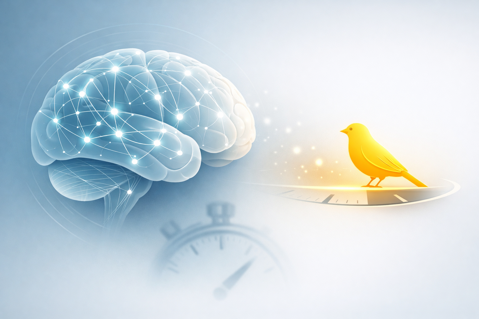
The canary doesn’t tell miners when the mine will collapse. It tells them that conditions are changing—and that attention is required. Reaction time, in this sense, isn’t just slowing. It’s signaling.
References
Du Y, Li R, Xing Y, Calhoun VD. Joint Aging Patterns in Brain Function and Structure Revealed Using 27,793 Samples. Research 2025;8:Article 0887. https://doi.org/10.34133/research.0887
Reuter-Lorenz PA, Cappell KA. Neurocognitive aging and the compensation hypothesis. Current Directions in Psychological Science 2008;17(3):177–182.
Buckner RL. The cerebellum and cognitive function: 25 years of insight. Neuron 2013;80(3):807–815.
Salthouse TA. The processing-speed theory of adult age differences in cognition. Psychological Review 1996;103(3):403–428.
Bernard JA, Seidler RD. Moving forward: Age effects on the cerebellum underlie cognitive and motor declines. Neuroscience & Biobehavioral Reviews 2014;42:193–207.
Arleo A, Bareš M, Bernard JA, et al. Consensus paper: cerebellum and ageing. Cerebellum 2024;23:802–832.


Leave a Reply